


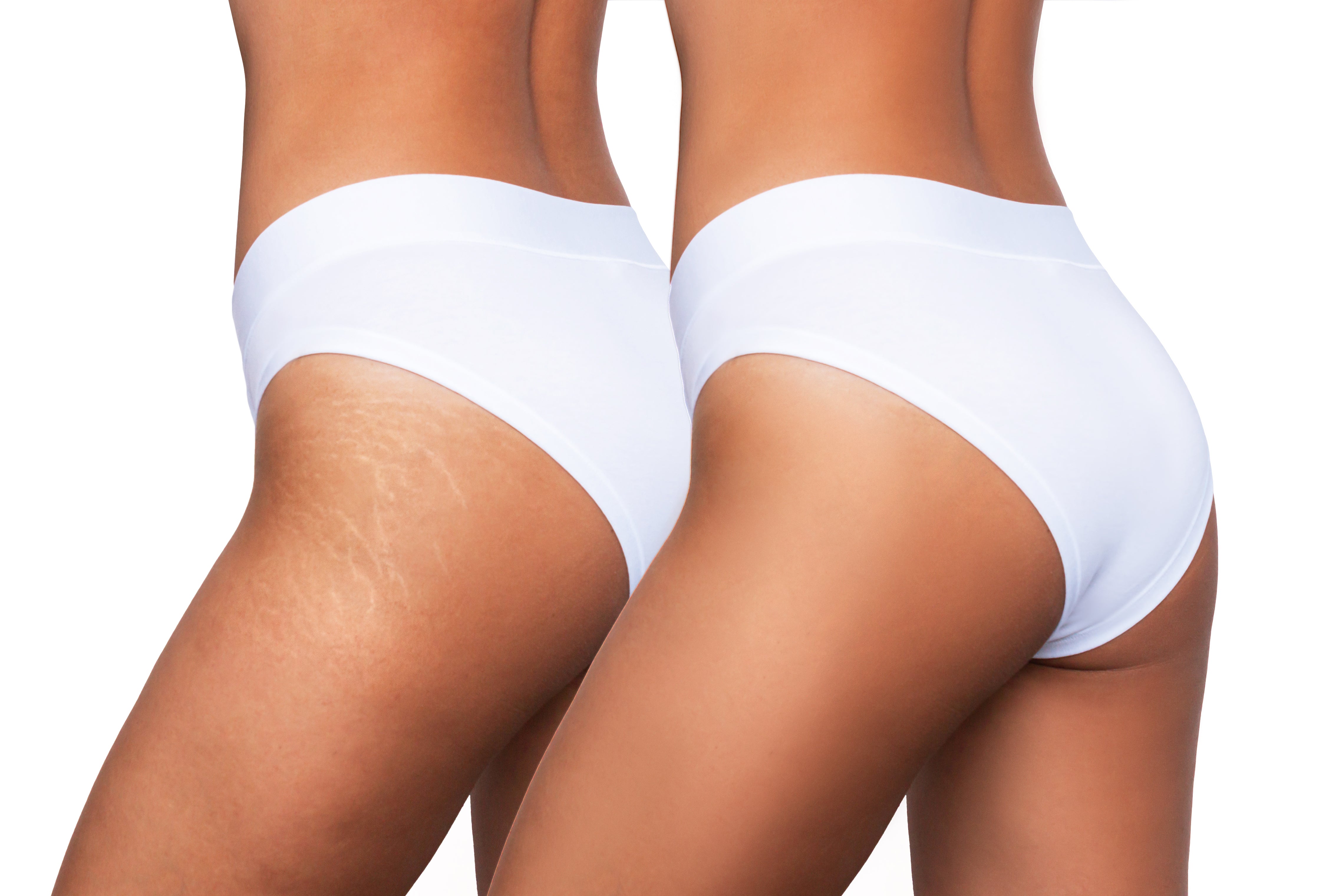


Developmental skin differences involving vessels or pigment, present at birth or noticed in early childhood.
Birthmarks & Moles
Most are harmless. Some are not.
Birthmarks and moles arise from variations in blood vessels or pigment cells during skin development. We approach them through precise diagnosis, risk assessment and evidence-based treatment selection — rather than cosmetic removal alone.

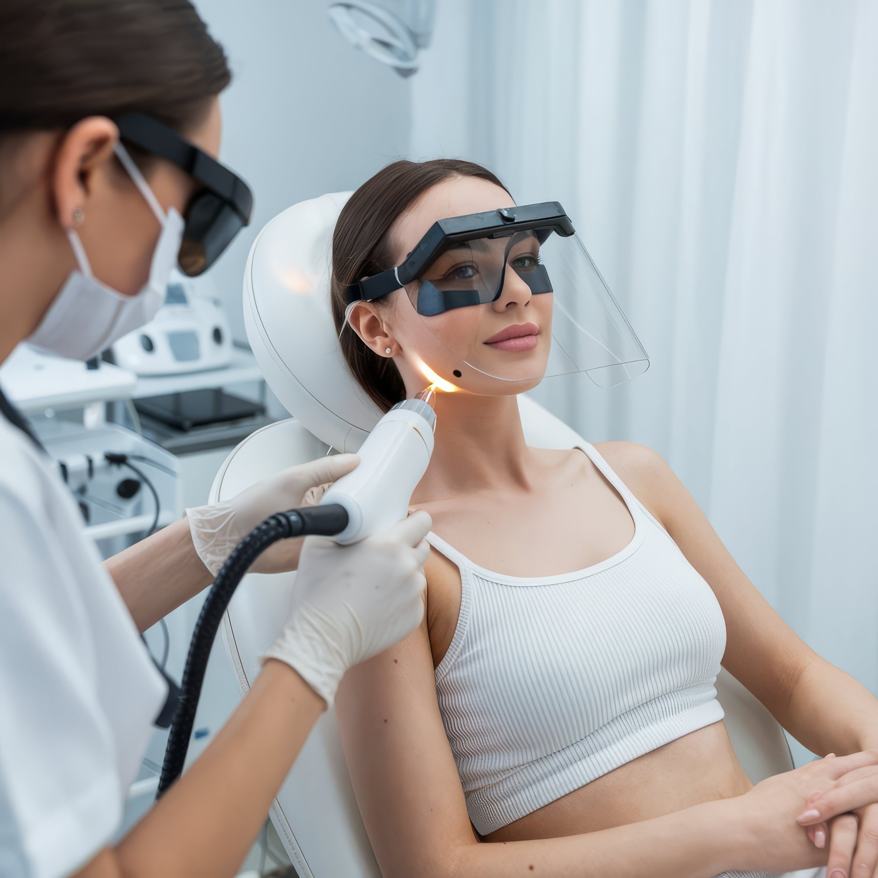
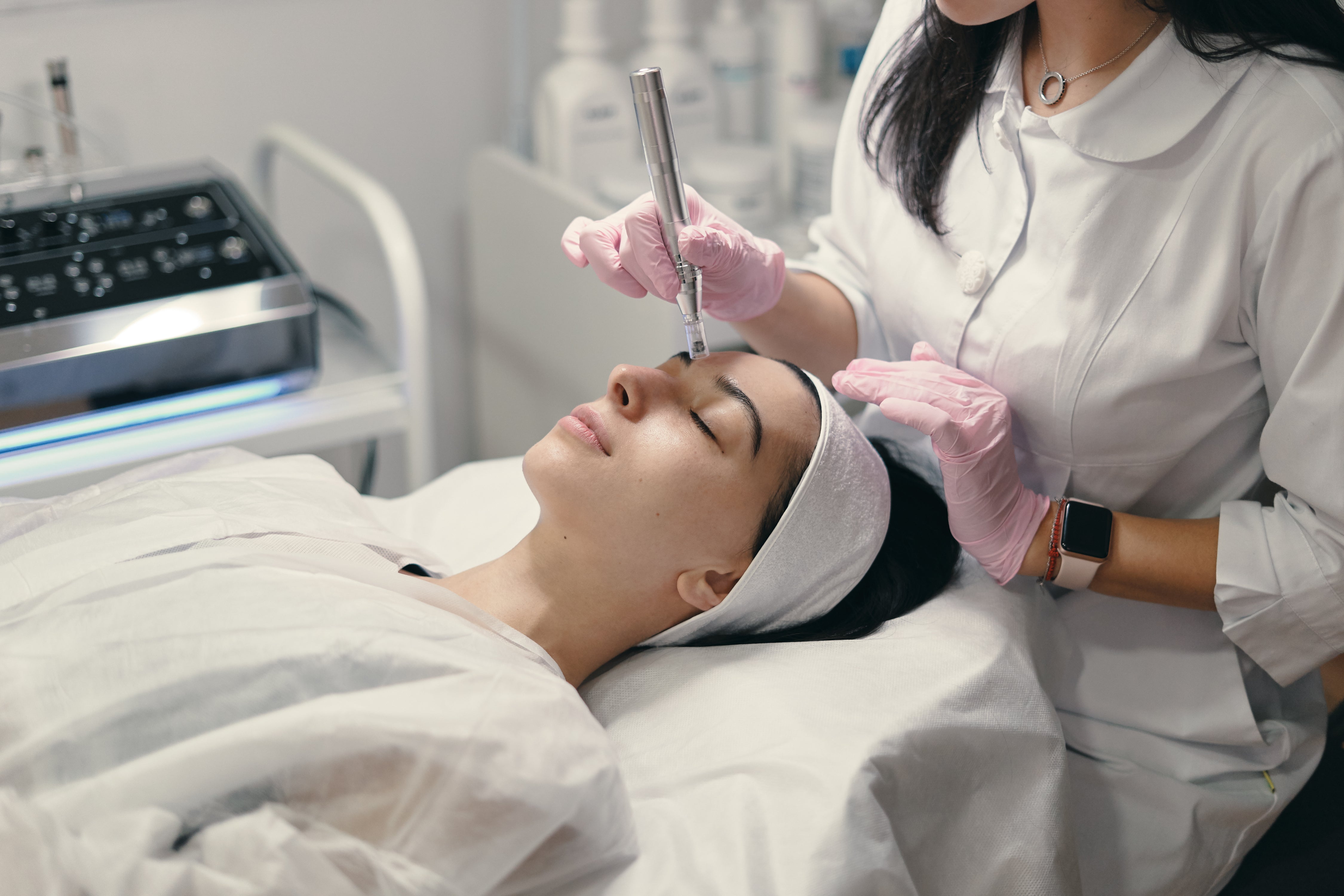
Diagnosis first, then the laser
Some lesions need a biopsy, not a device. Knowing which is the entire job.
At birthwhen most birthmarks appear
ABCDEthe change checklist for moles
IV–VIFitzpatrick types we tune for

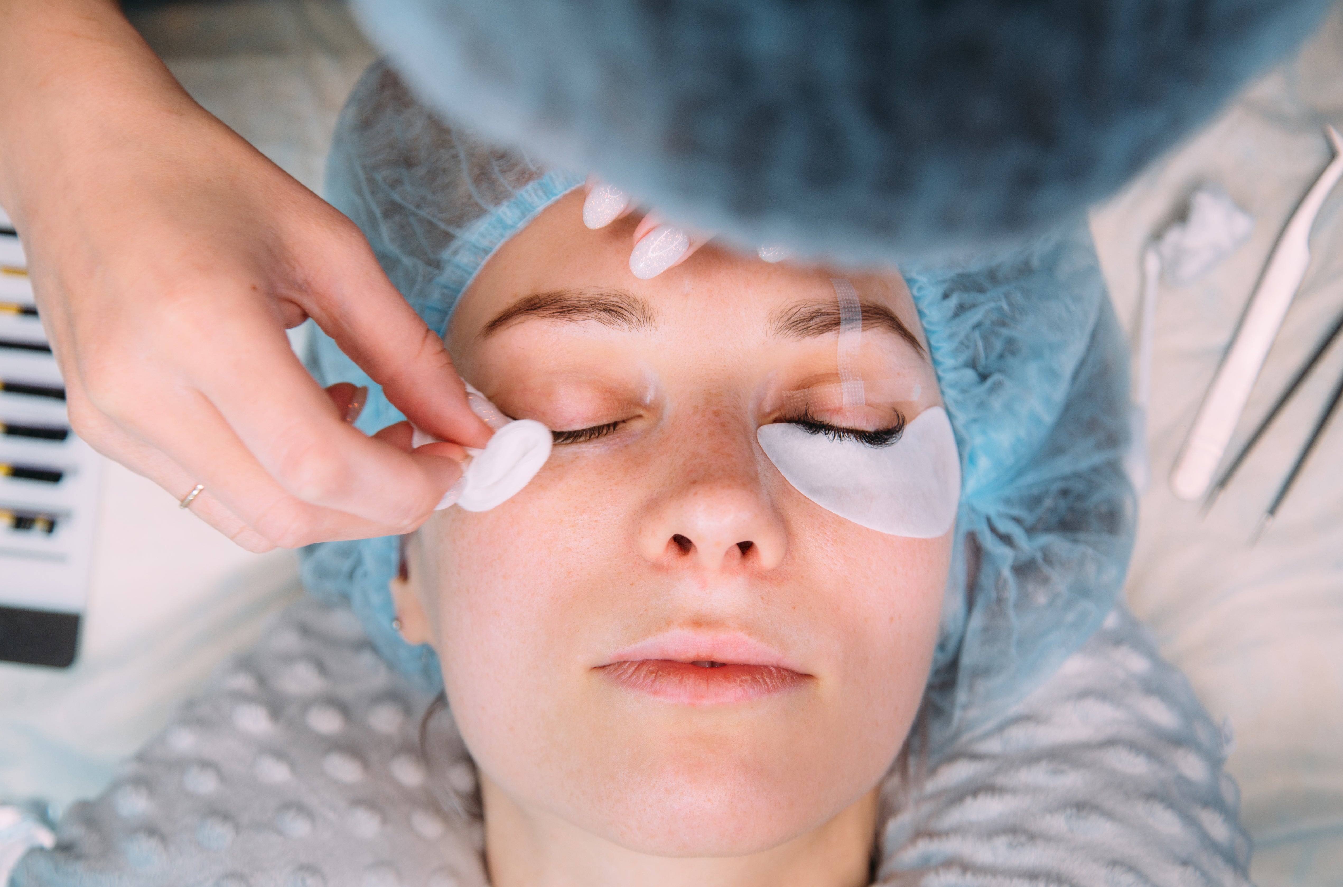
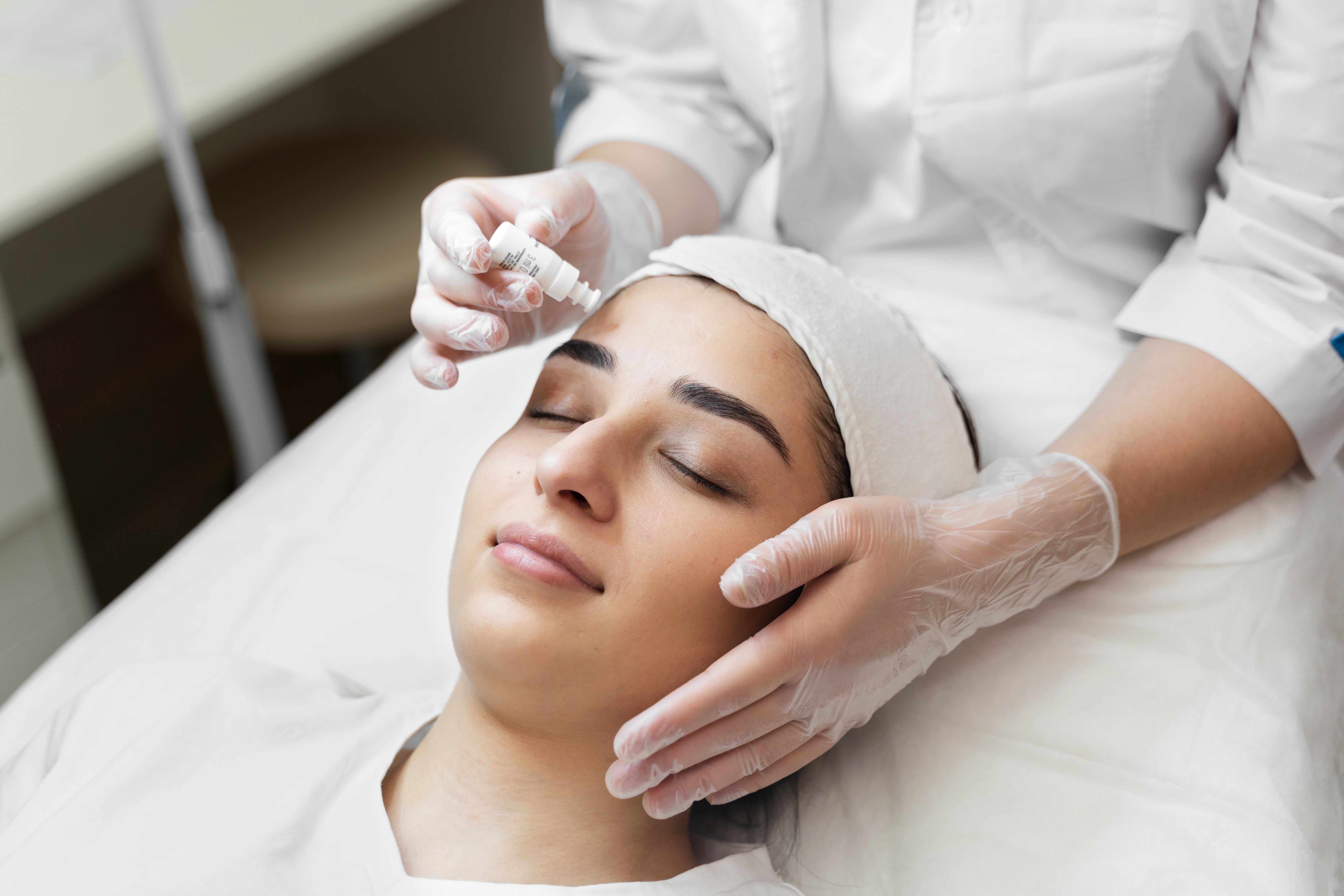
Why Allodermis
The difference is in the doctor
Removing a lesion is easy. Knowing which lesion should never be removed with a laser is the part that needs a dermatologist.
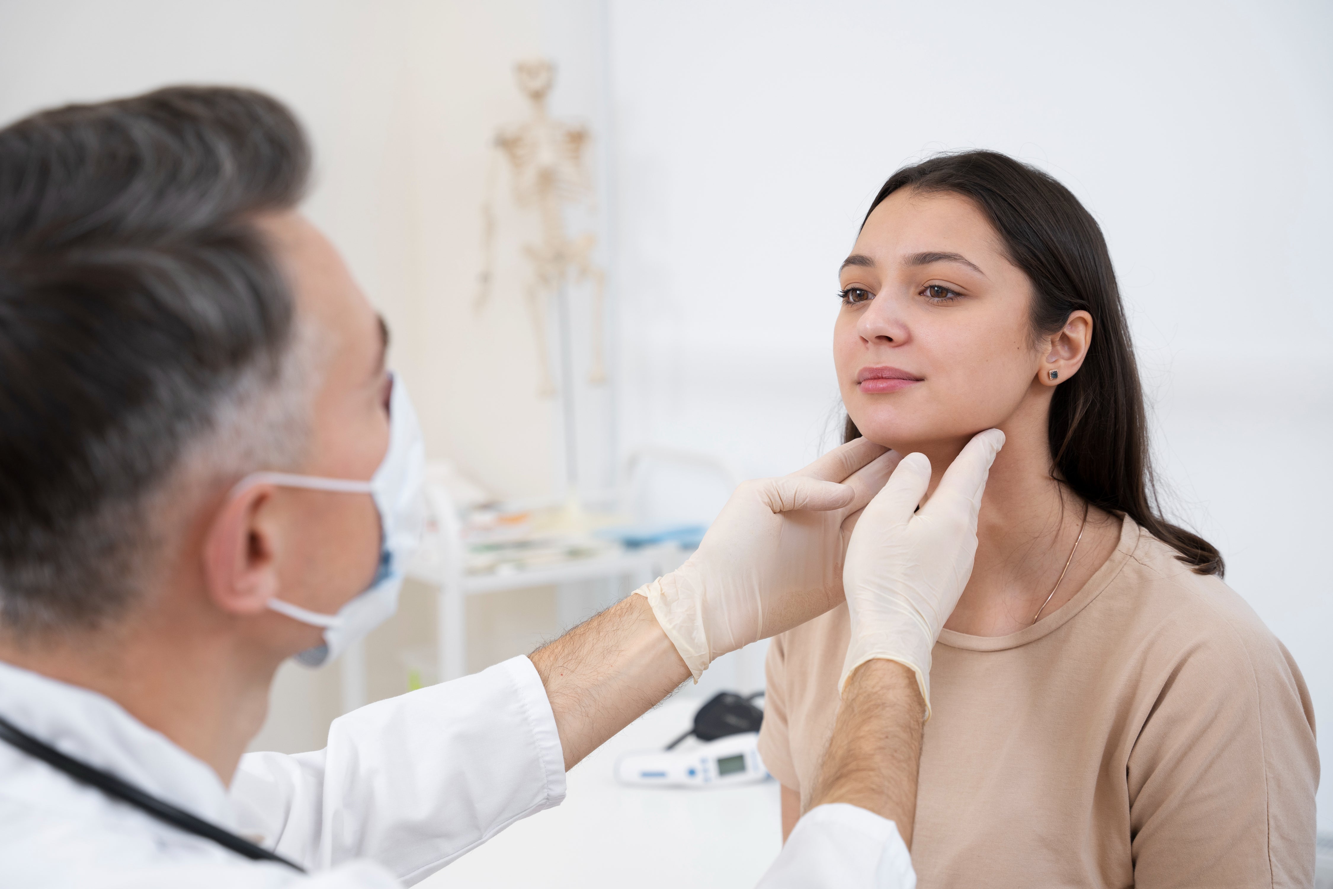
- AIIMS MD DermatologistsEvery assessment is made and supervised by MD dermatologists trained at AIIMS New Delhi — not technicians or salons.
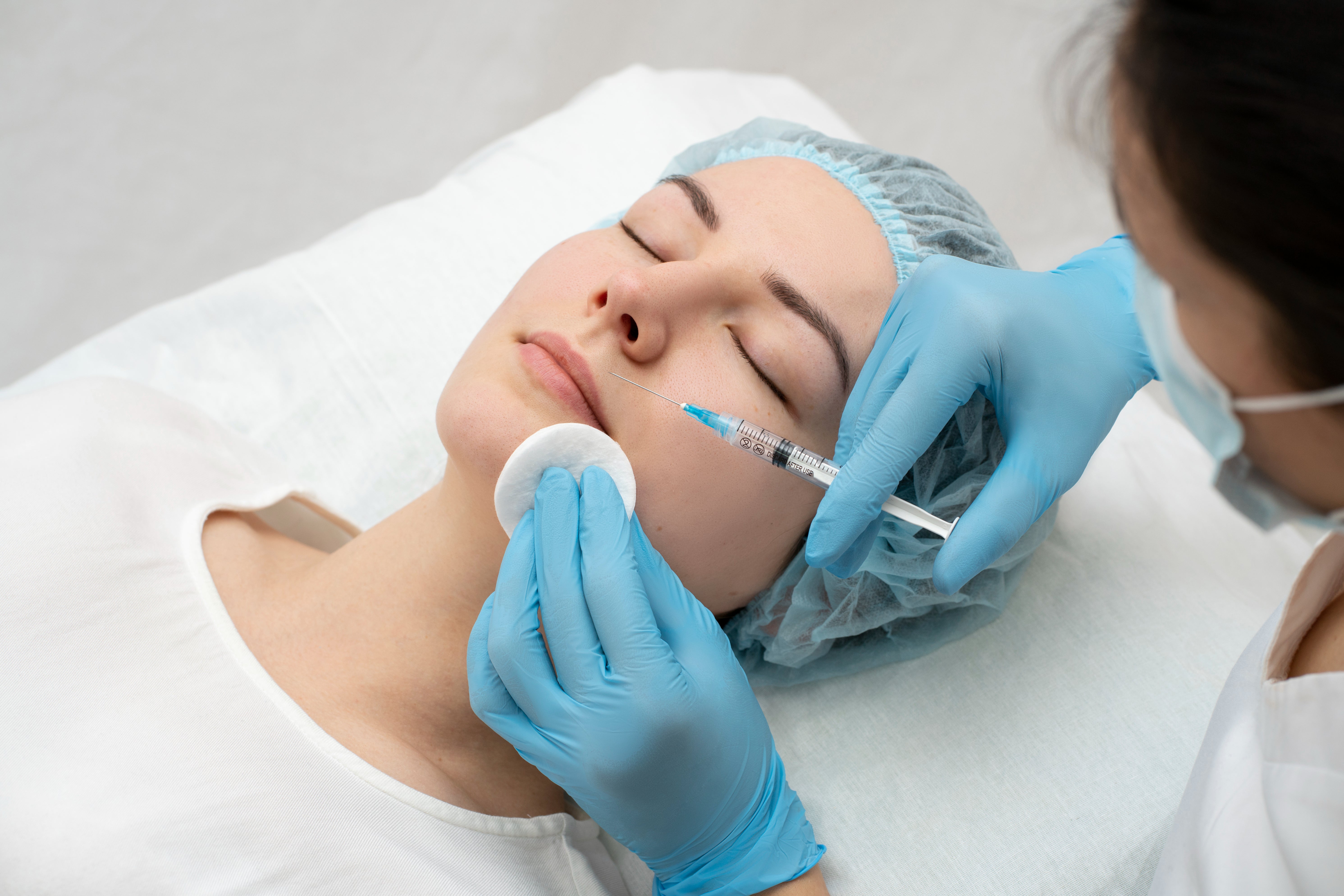
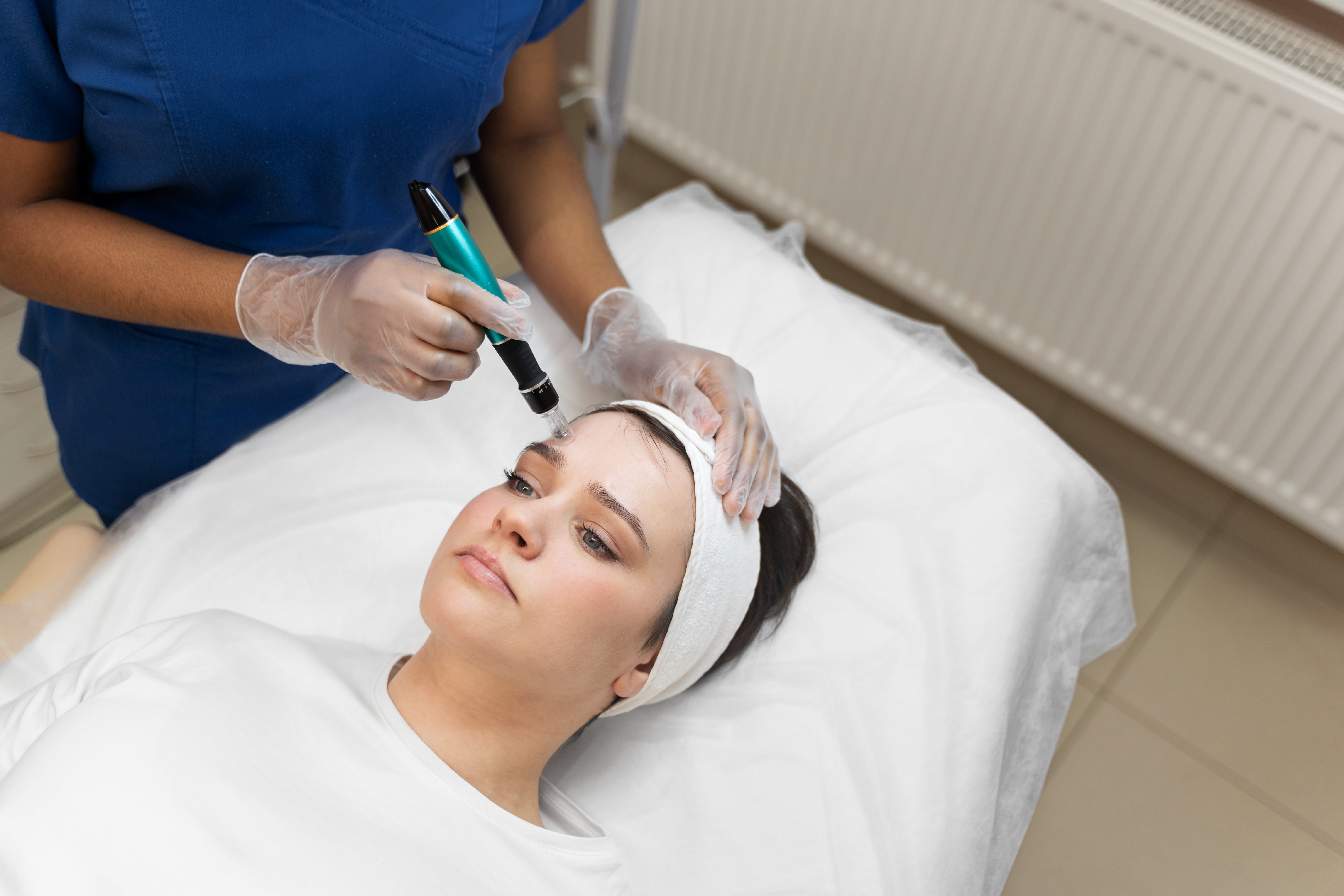
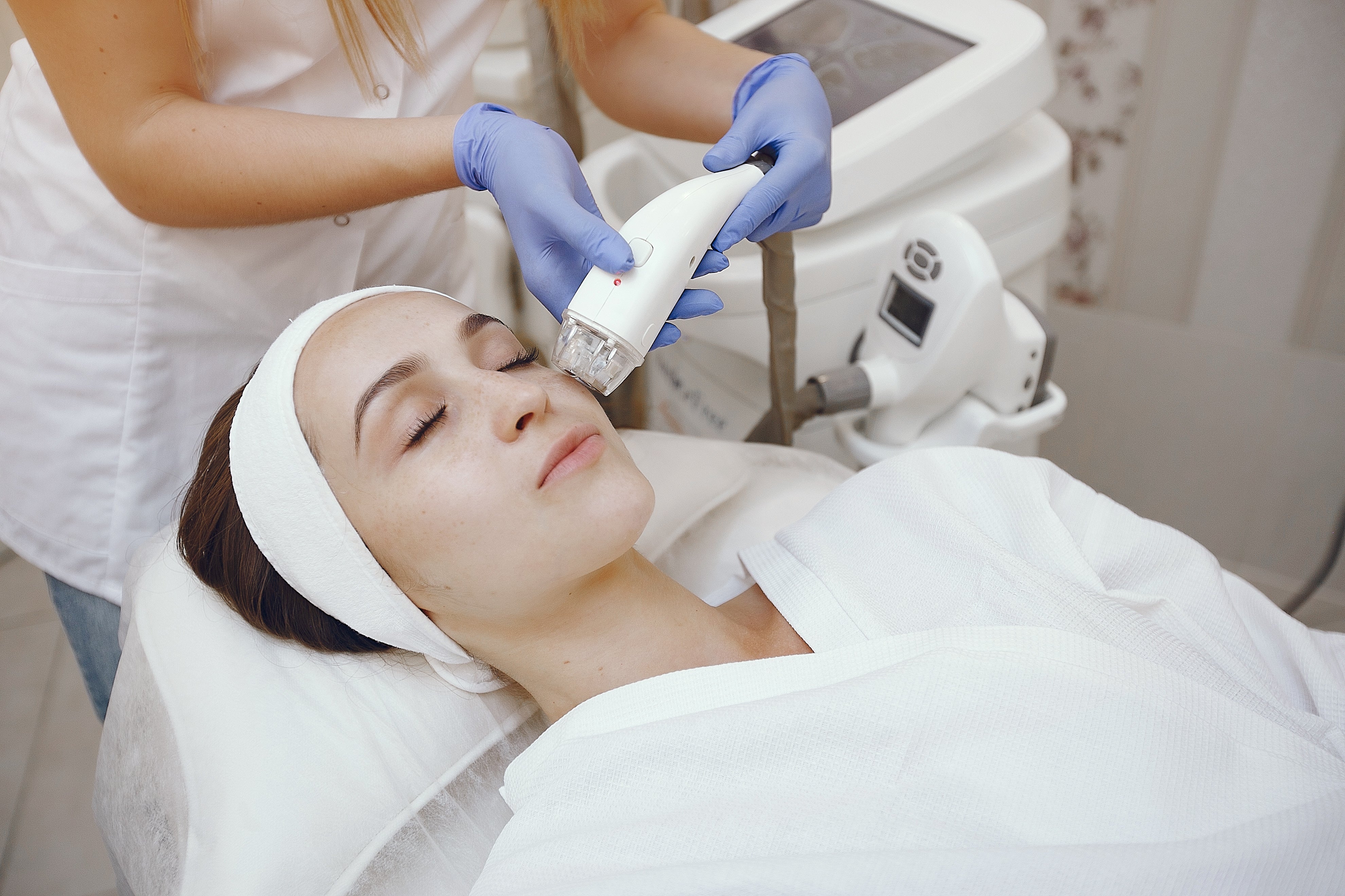
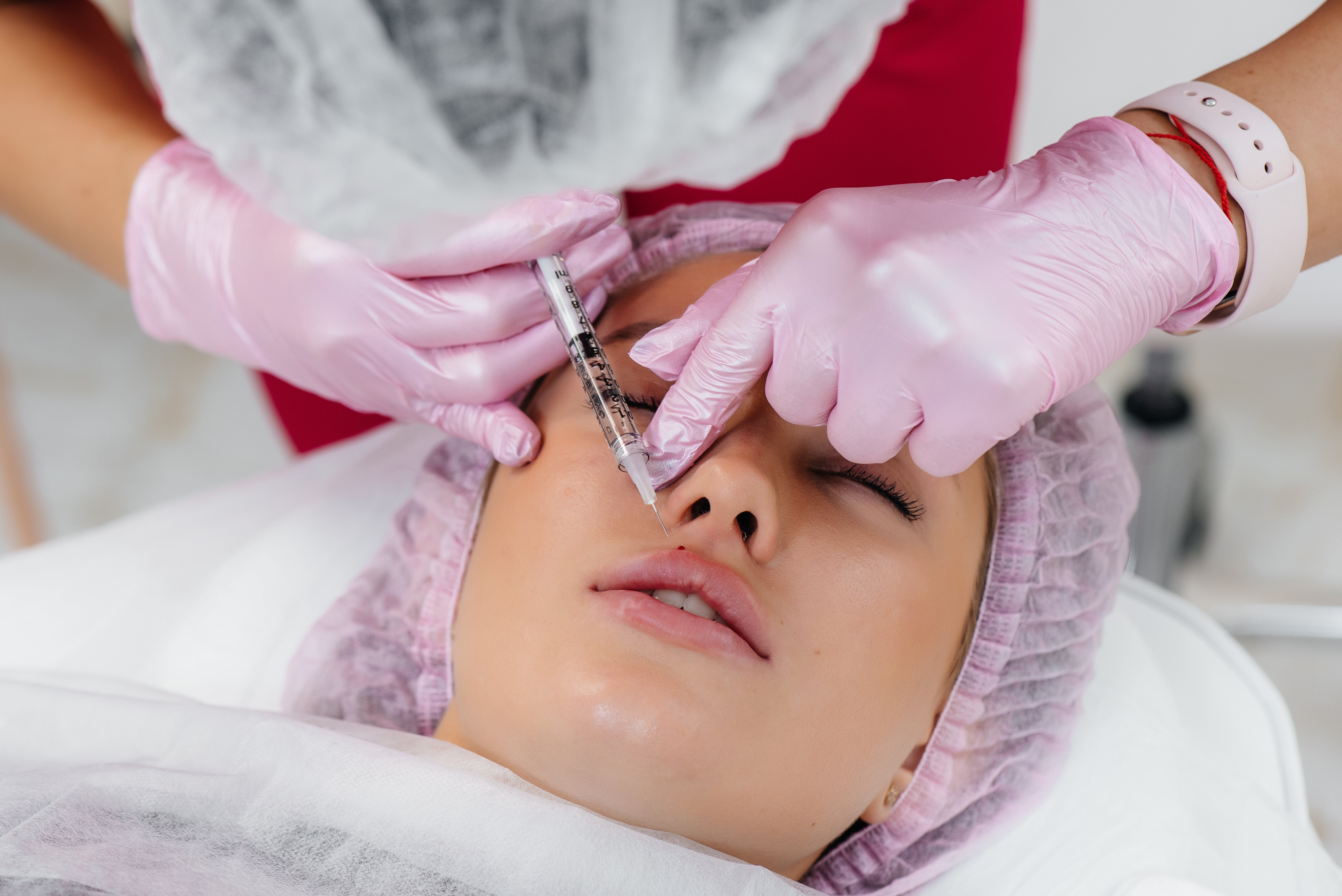
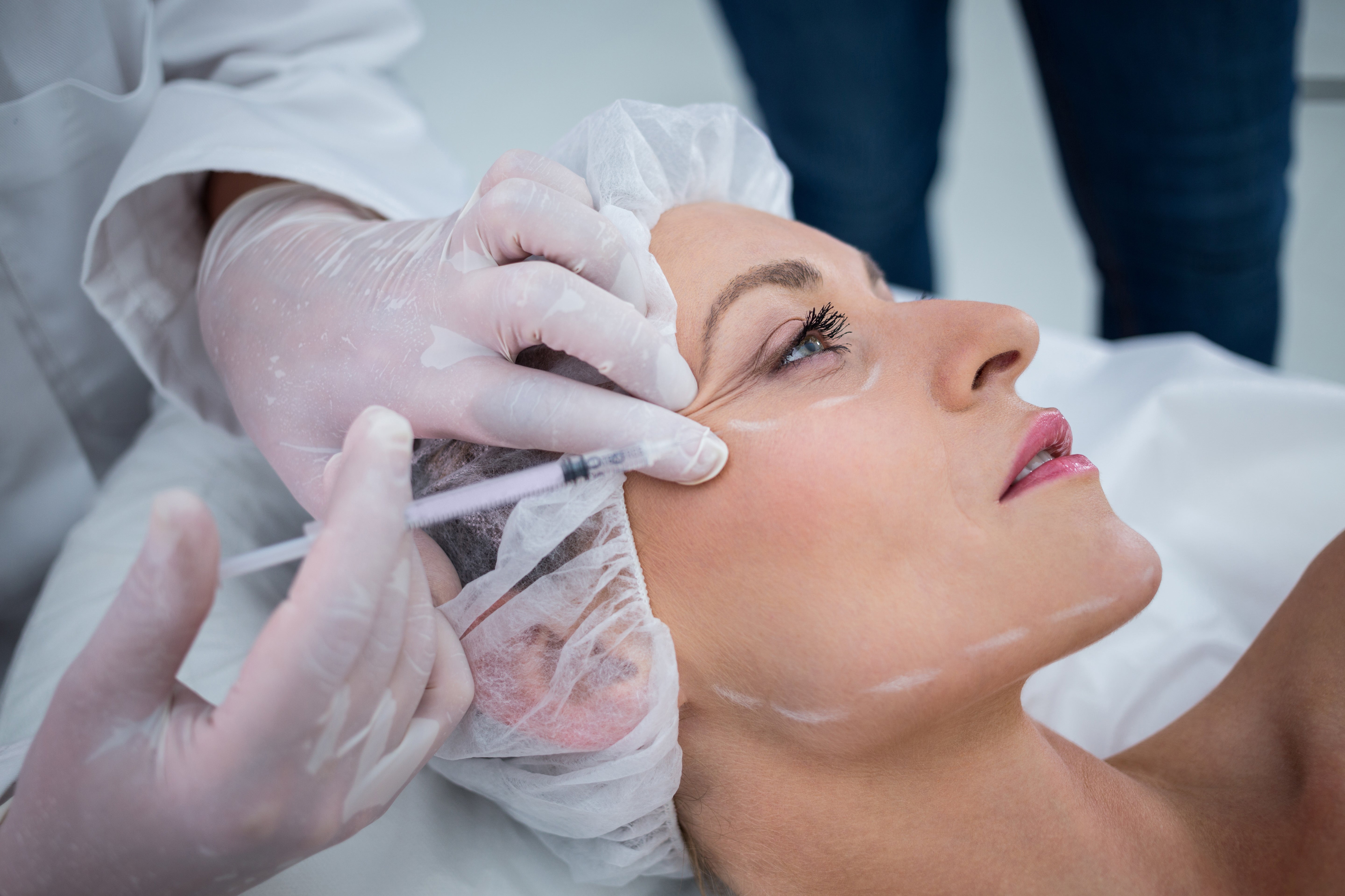
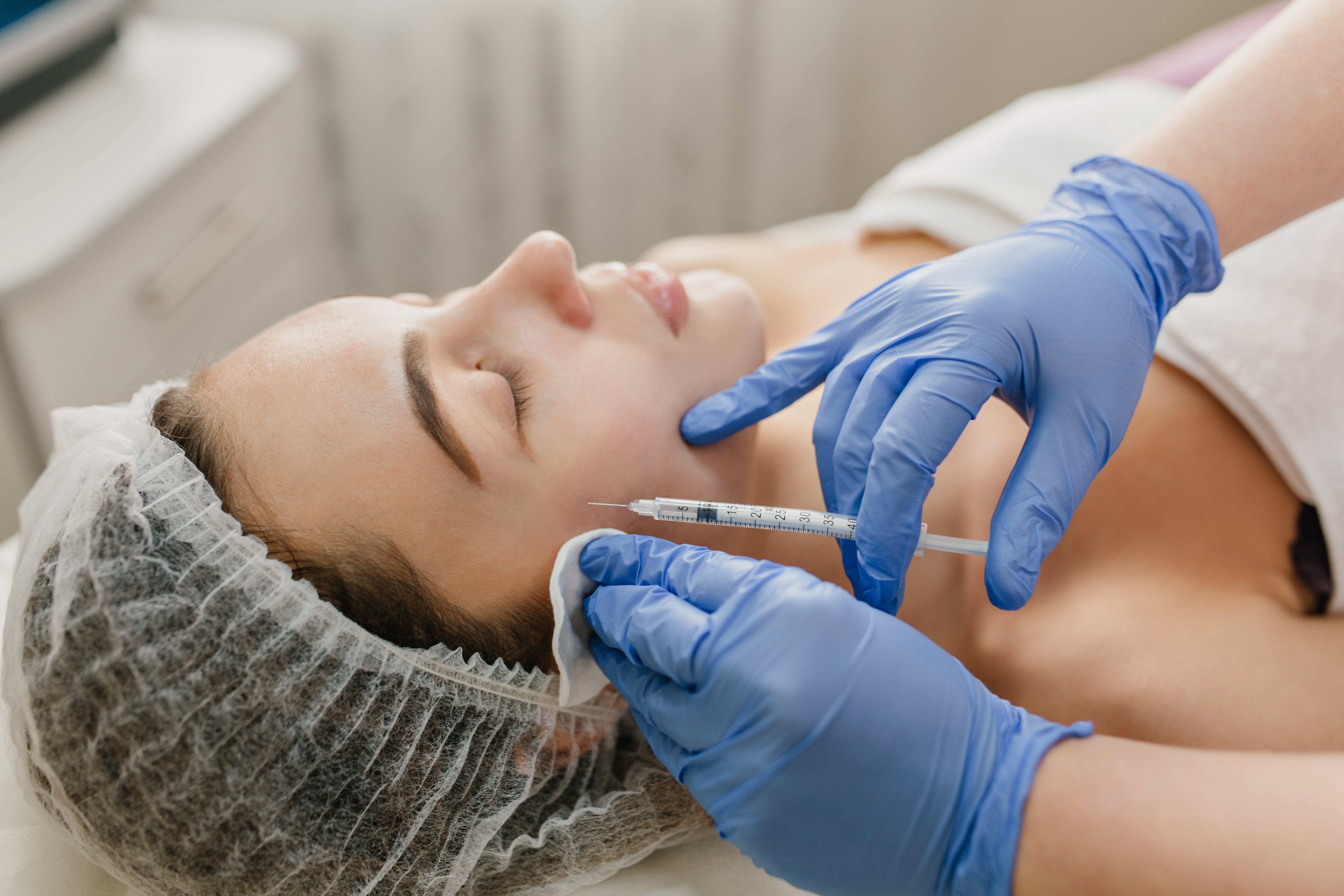
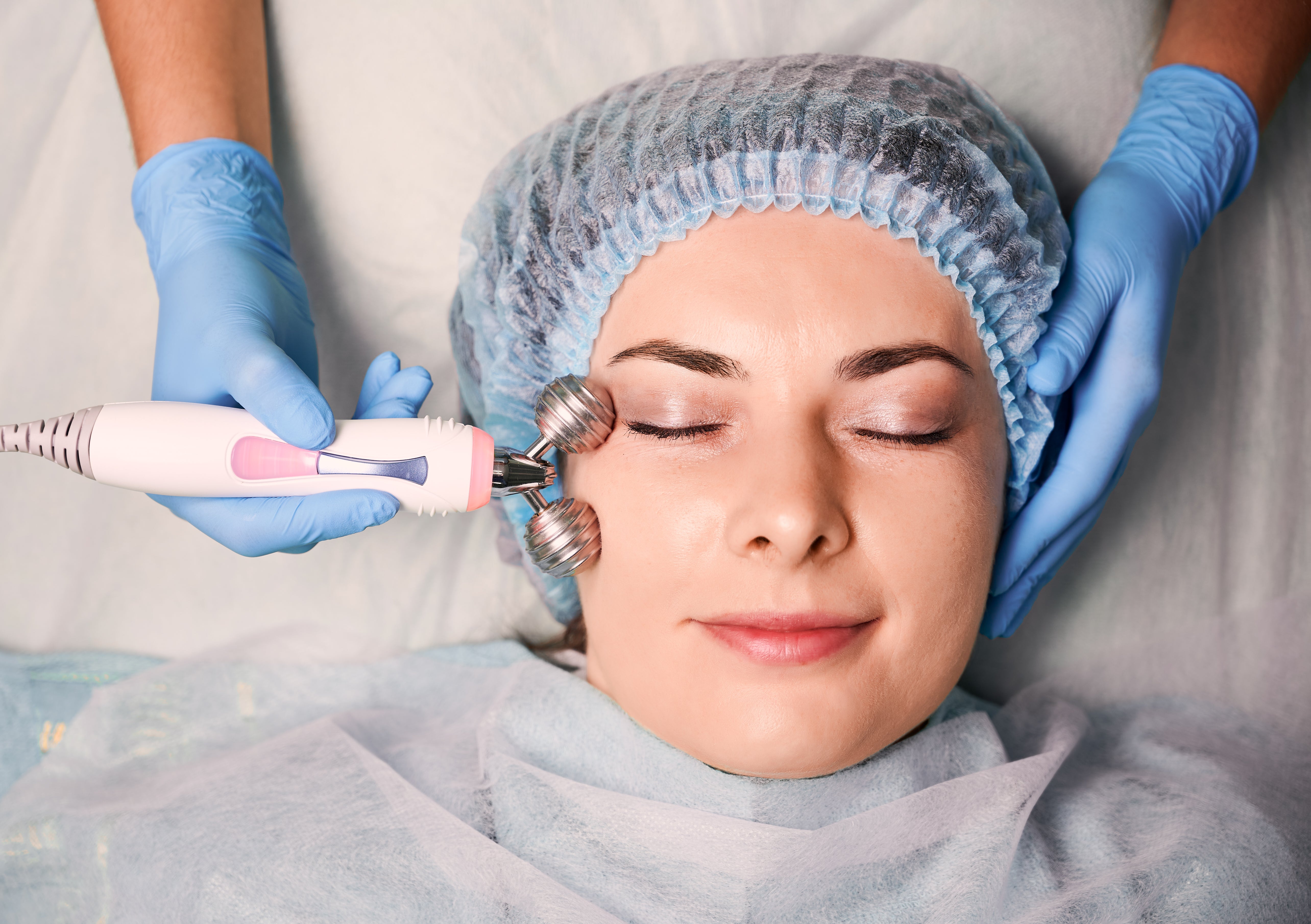
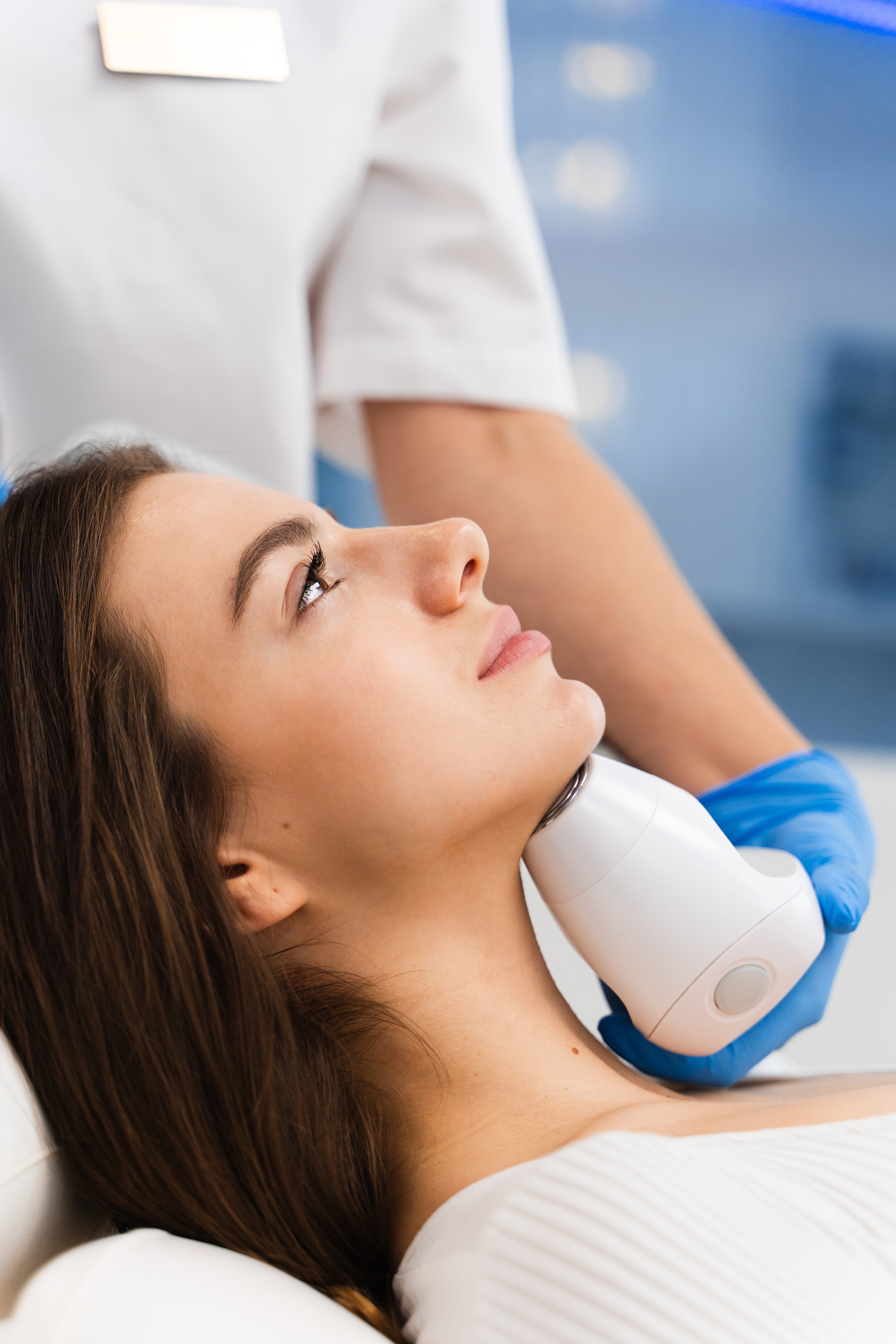
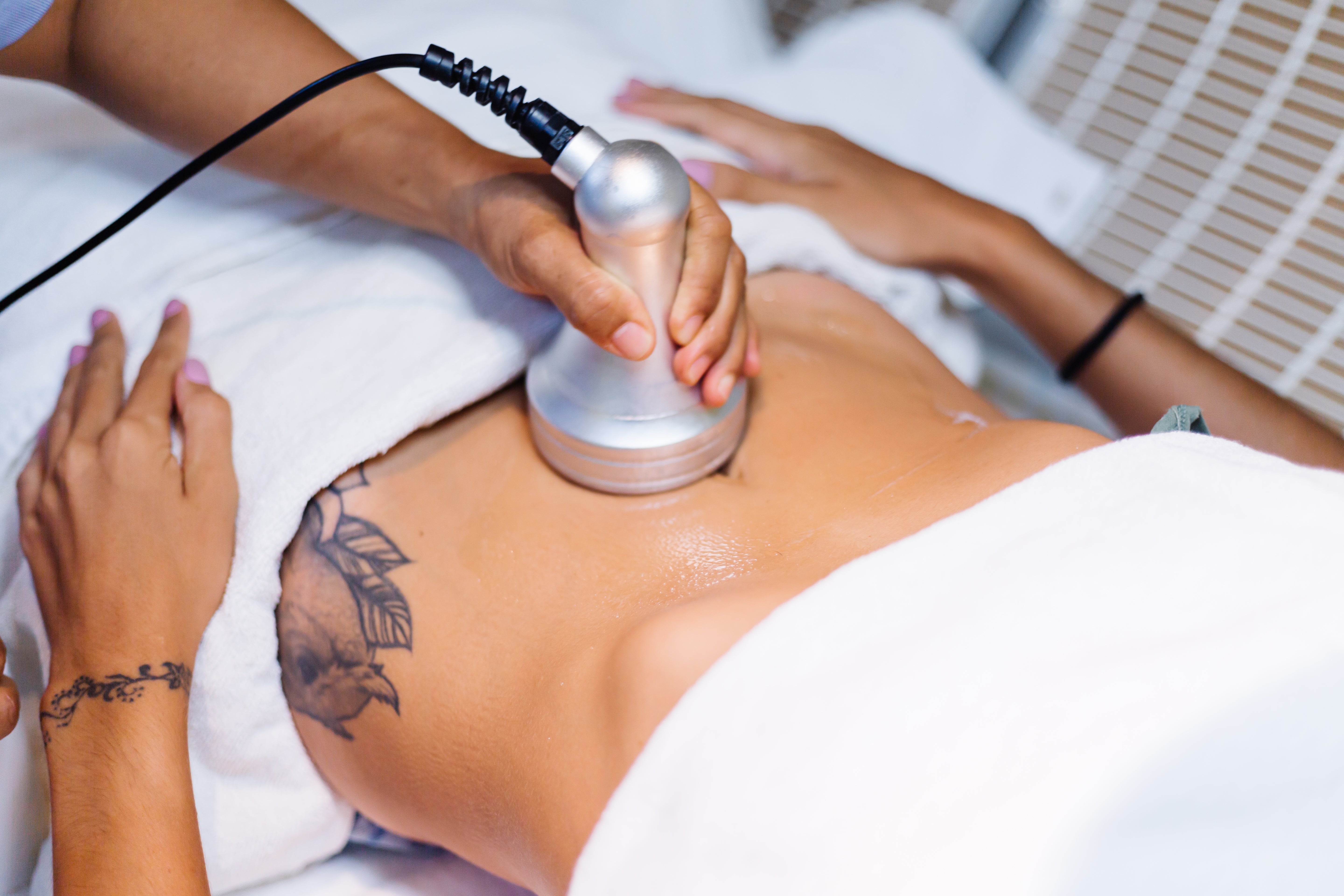
- Evidence-based technologyPulsed dye, Q-switched Nd:YAG and radiofrequency platforms, used strictly within USFDA-approved, peer-reviewed protocols.
- Tuned for Indian skinWavelength, fluence and spacing adjusted for Fitzpatrick IV–VI skin, where the risk of post-laser pigment change is real.
- Diagnosis before removalWe establish the lesion type and its risk profile first, then decide whether it needs treatment, monitoring or a biopsy.
01 · The Basics
What they actually are
Two different mechanisms sit behind almost everything on this page. Separating them is the first thing an assessment does.
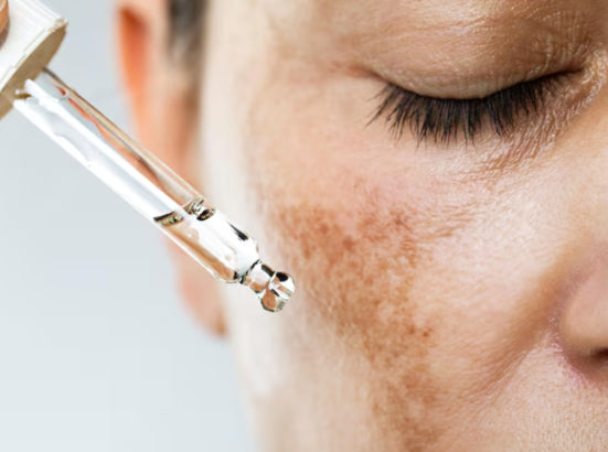
Localized clusters of melanocytes, the pigment cells. Most are benign, but change-based warning signs need medical review.
iVascular or pigment variation during skin development
iiMoles are localized clusters of melanocytes
iiiThe large majority of both are benign
ivA minority need monitoring or intervention
vRisk is read from type, size and change
02 · Types
Two families, two mechanisms
Which family a lesion belongs to decides the entire treatment path — the device, the drug, and whether either is appropriate at all.
Vascular birthmarks
Caused by abnormal blood vessel development
- Nevus simplexLight pink patches that often fade over time
- Port wine stainPersistent red to purple patches that may thicken with age
- Infantile hemangiomaGrows after birth, then gradually shrinks. Some need treatment
Pigmented birthmarks & moles
Caused by melanocyte activity
- Congenital melanocytic nevusPresent at birth. Risk depends on size
- Cafe au lait maculesLight brown patches. Multiple lesions may need evaluation
- Dermal melanocytosisBluish grey patches, common in infants, often fade
Not sure which one you're looking at?
Colour alone does not settle it. Depth, borders, history and rate of change do — which is exactly what a dermatological assessment separates before anything is treated.
- AIIMS MD Dermatologists
- USFDA-approved technology
- Tuned for Fitzpatrick IV–VI
- Diagnosis before removal
03 · Root Cause
Why they form
Most cases are sporadic. Genetics and developmental factors set the pattern — nothing a parent did or did not do causes them.
Capillary developmentVessels form and persist in a localized area during fetal growth instead of regressing.
Post-birth growth phaseHemangiomas proliferate after birth, then involute slowly over years.
Depth and flowHow deep the vessels sit governs colour, thickening and laser response.
Sporadic occurrenceThe great majority arise without any inherited or preventable cause.
Melanocyte clusteringPigment cells group together in a defined area rather than spreading evenly.
Layer of pigmentEpidermal versus dermal placement changes both the colour seen and how treatable it is.
Genetic & developmental factorsInfluence the number, size and distribution of lesions.
Size at birthIn congenital melanocytic nevi, size is the main determinant of long-term risk.
The Core Principle
A mole that needs pathology will never be a laser case.
✕A laser removes the lesion, not the diagnosisThe mark disappears and the answer disappears with it. Nothing is left to examine, and nothing is confirmed.
✓Removal without histopathology removes the evidenceWhere tissue needs reading, excision keeps the specimen intact so the lesion is both treated and understood.
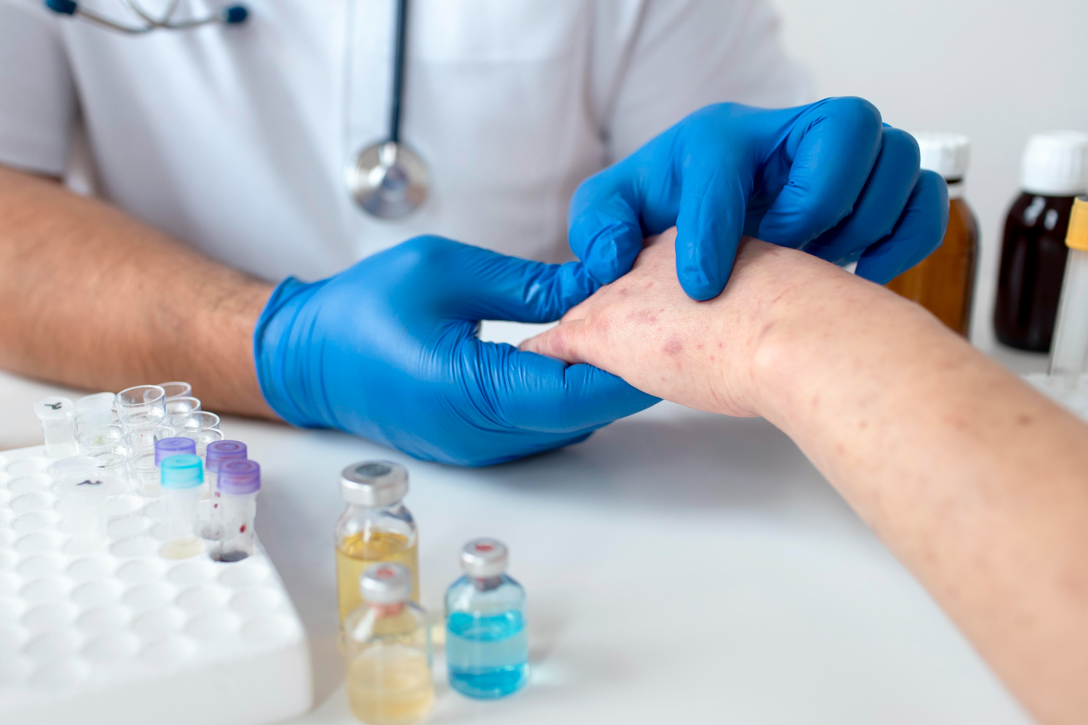
04 · Red Flags
When to refer urgently
None of these mean cancer on their own. They mean the lesion needs to be looked at properly, now — immediate dermatology evaluation rather than a wait-and-watch.
Speak to a dermatologist today→- Rapid growth or change in the lesion
- Irregular borders or multiple colours
- Bleeding, ulceration or pain
- Large congenital moles
- More than six cafe au lait spots in a child
The ABCDE check. A mole should be reviewed urgently if it shows any of these five changes.
AAsymmetry
BBorder irregularity
CColour variation
DDiameter increase
EEvolving
05 · Treatment Principles
We treat only when it is indicated
Medically or cosmetically indicated, and only once the lesion type is established. The tool follows the diagnosis, never the other way round.
Vessel-targeted care
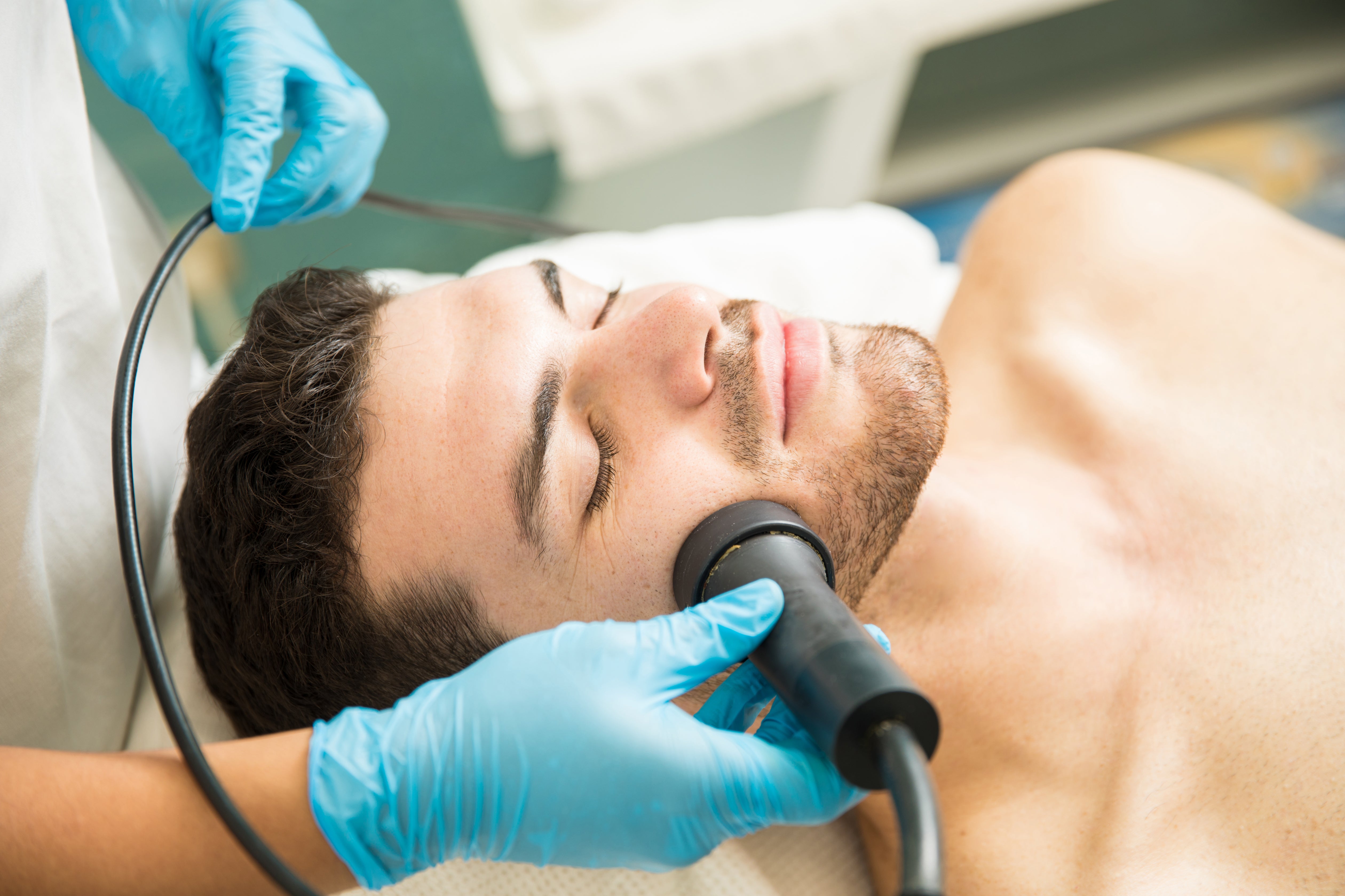
01Pulsed Dye Laser
The first-line option for port wine stains. Targets haemoglobin inside the abnormal vessels while sparing the surrounding skin.
Best for port wine stainsCourse multiple sessions
02Oral Propranolol
Medical therapy for infantile hemangiomas. High-risk cases benefit from early evaluation, and treatment is most effective when started early under supervision.
Best for infantile hemangiomaStart early, supervised
03Targeted Procedures
Case-specific interventions for residual lesions after a primary treatment course, planned individually rather than sold as a package.
Best for residual lesionsPlan case by case
Pigment-targeted care
04Q-Switched Nd:YAG Laser
Selective use in suitable pigmented lesions. Settings are chosen for darker skin types, where post-inflammatory pigmentation is the main risk.
Best for benign hypermelanosisChoice by pigment depth
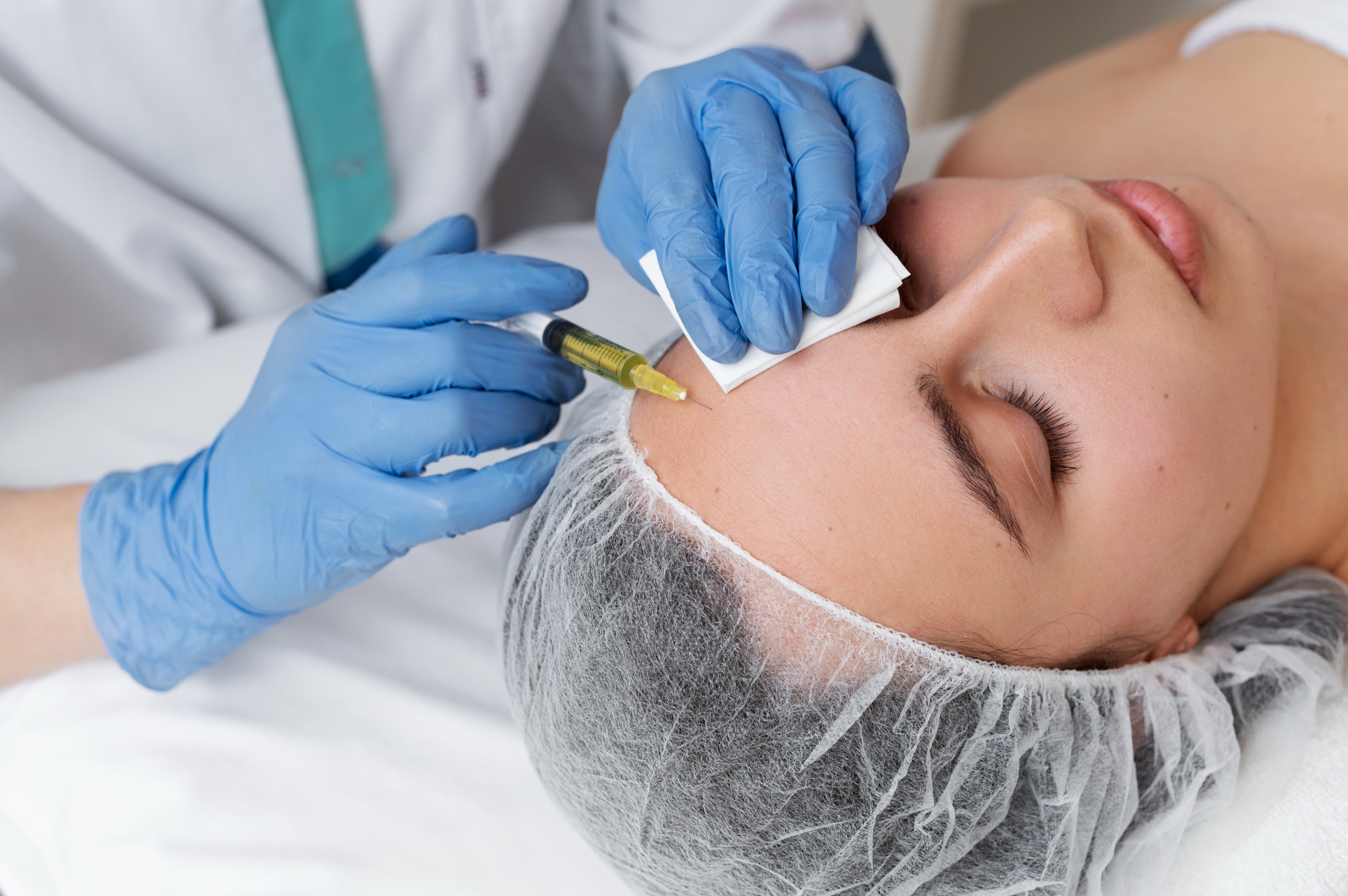
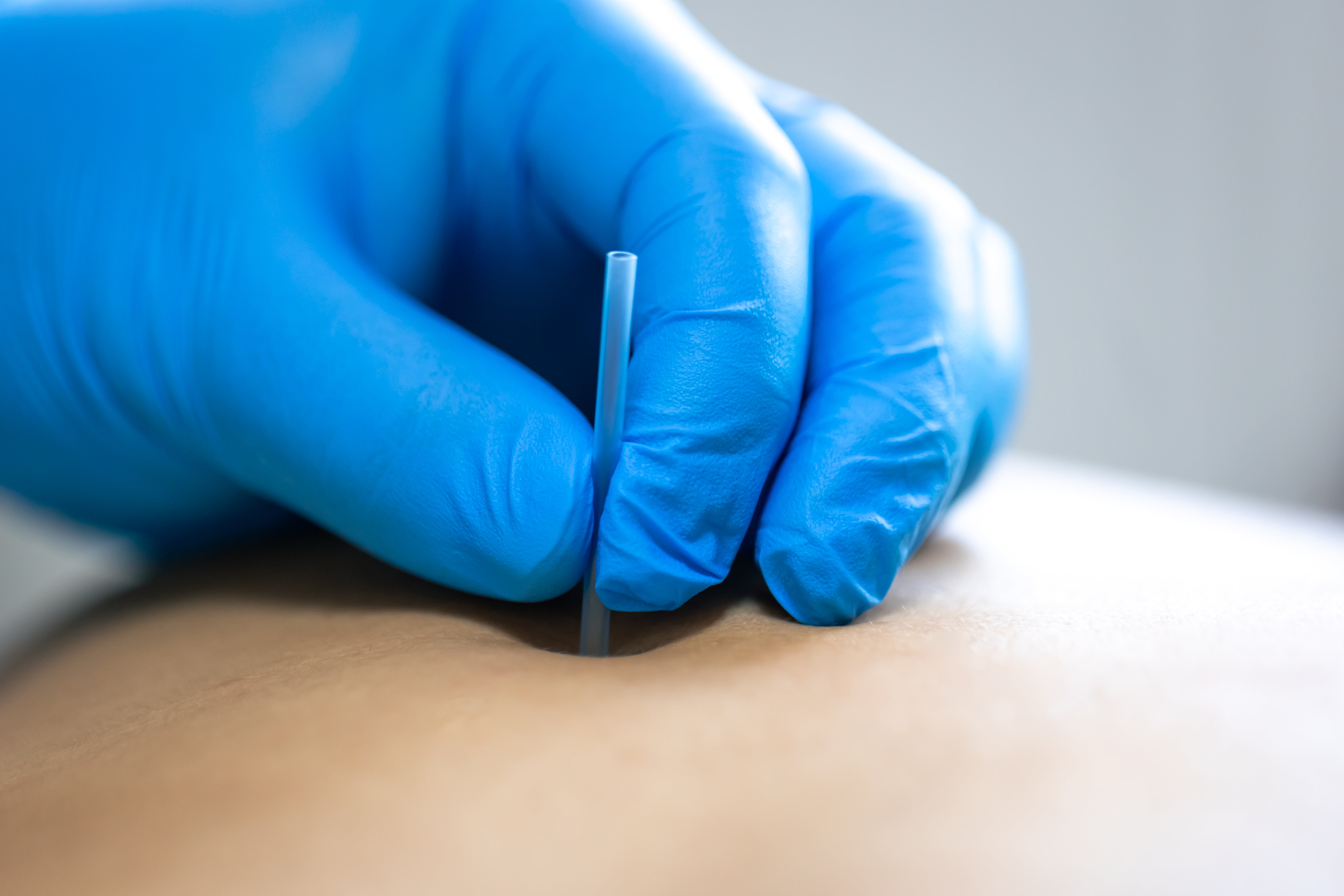
05Excision & Histopathology
Mole removal is preferred wherever a tissue diagnosis is required. Surgical removal preserves the specimen so the lesion can actually be read under a microscope.
Best for changing or atypical molesGives a tissue diagnosis
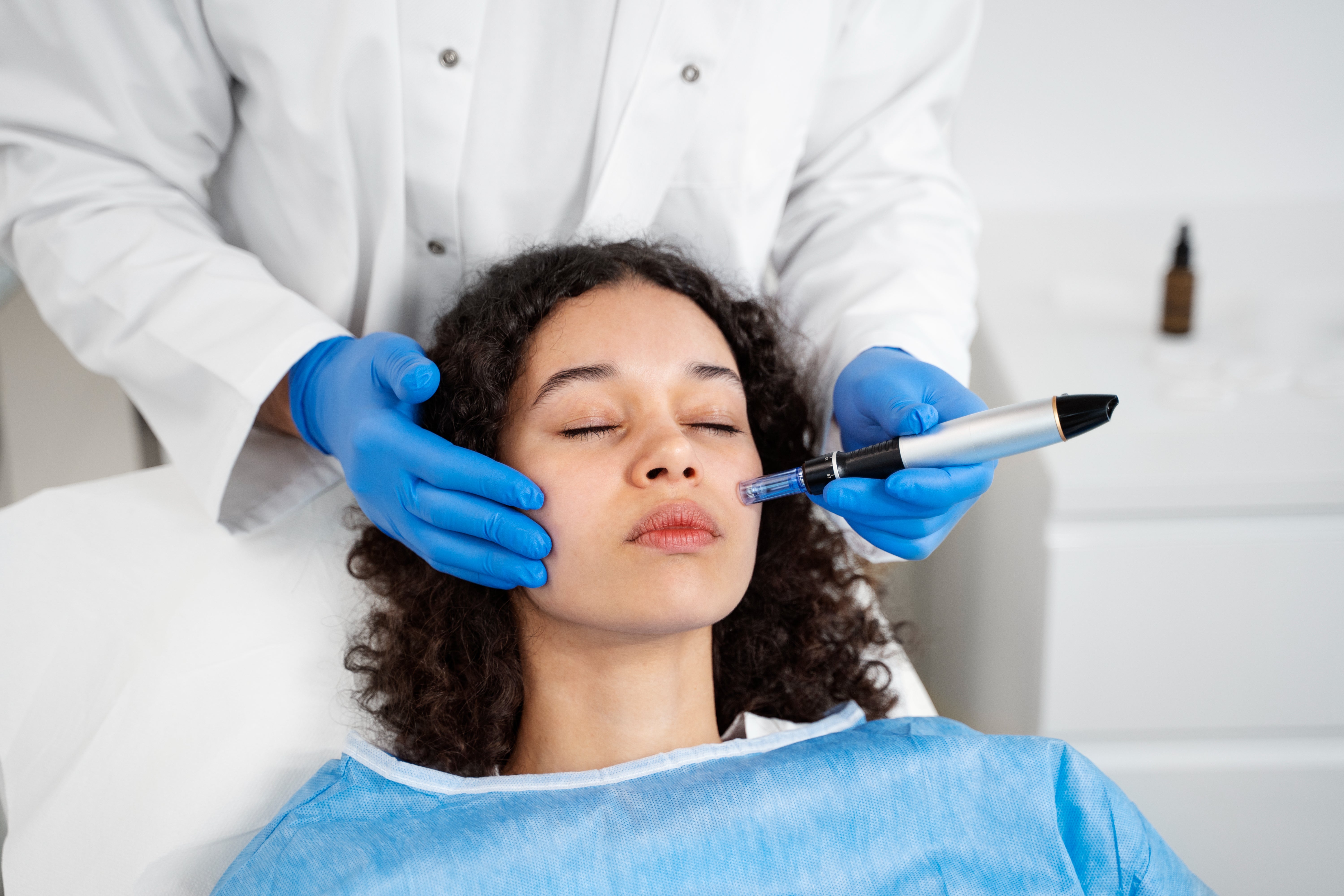
06Radiofrequency Ablation
Precise removal of small, clearly benign raised lesions, with controlled depth and minimal thermal spread into the surrounding skin.
Best for small benign raised lesionsRequires a confirmed diagnosis
Important
We do not recommend blanket laser removal for all moles. A proper diagnosis is essential before treatment, and some lesions require a biopsy instead of a laser.
06 · Typical Pathways
How a case actually runs
Every path opens with diagnosis. What follows depends on the family the lesion belongs to and what the assessment finds.
01Port wine stain
Diagnosis & mapping→Pulsed dye laser course→Sun protection & review
02Infantile hemangioma
Risk assessment→Oral propranolol if indicated→Monitored involution
03Changing mole
Clinical & dermoscopic exam→Excision→Histopathology
04Benign pigmented patch
Confirm the diagnosis+Q-switched laser only if suitable
05Large congenital nevus
Size-based risk review→Long-term surveillance
07 · What to Expect
The honest version
Improvement here is real but partial, and it is earned across a course rather than in a single visit.
Multiple sessions
Most vascular and pigmented lesions need a planned series rather than one appointment.
01Strict sun protection
Sun protection is necessary during the course and after it, not optional.
02Improved, not always erased
Some lesions improve considerably but may not disappear completely.
03Long-term follow-up
Certain conditions need continued review well past the final session.
0408 · Diagnosis at Allodermis
The 5-step root cause protocol
Root cause diagnosis
We don't chase marks. We establish what the lesion is and what it is likely to do next — from hormones to nutrition to stress, addressed inside out.
Right products, only when needed
We don't believe in routines overloaded with products. We prescribe only what truly serves your skin and nothing more.
Dermatologists you can trust
MD Dermatologists from AIIMS New Delhi, trained in the science of skin and the art of empathy.
Science that serves you
Only USFDA-approved technologies and evidence-based protocols. No pseudoscience, no gimmicks. Just the truth that shows.
Inside-out wellness
Mind, body and skin treated as one ecosystem, because healthy skin isn't built in a clinic. It's built into your everyday life.
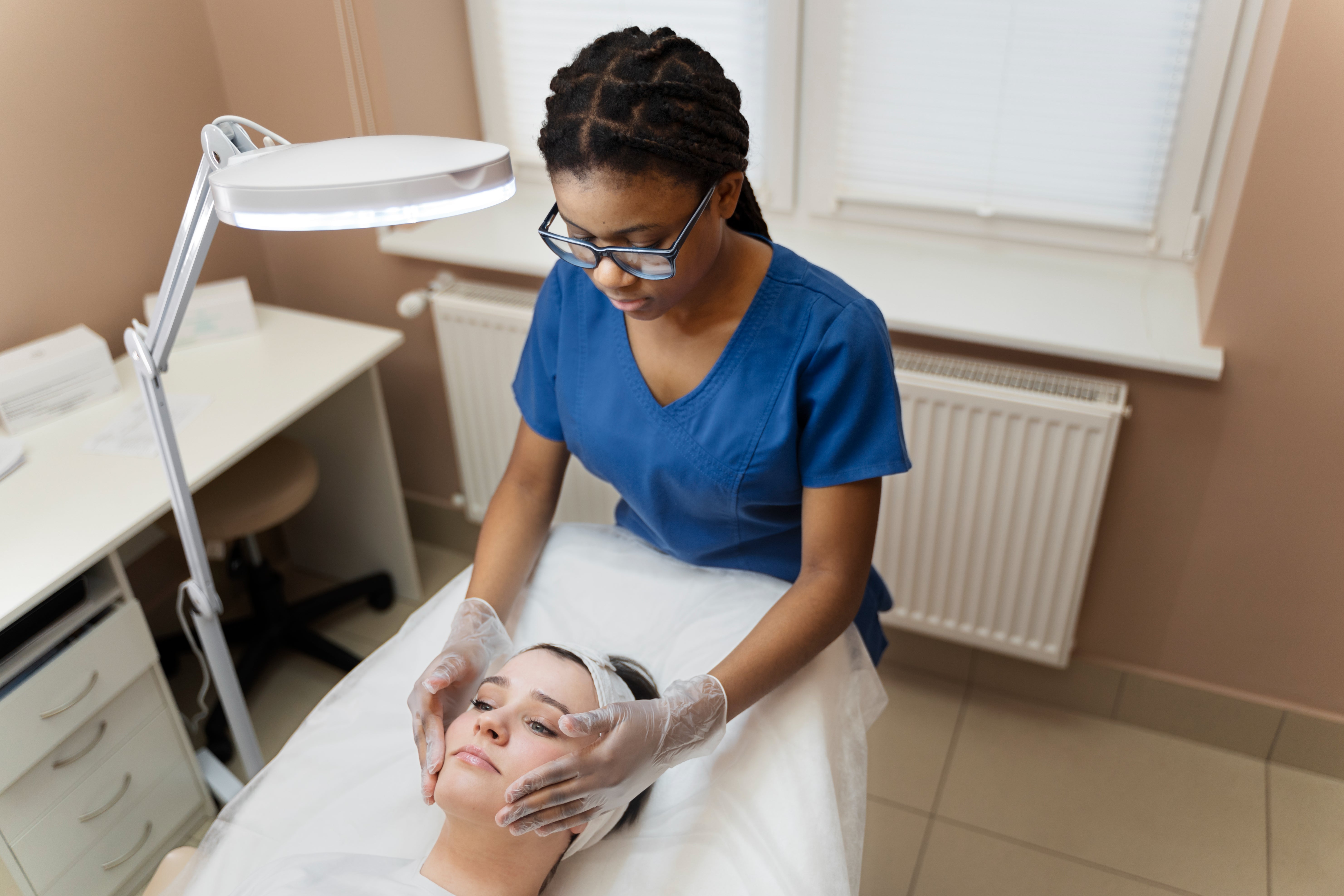
09 · Safety
Every case under medical supervision
Laser is safe for Indian skin when both the settings and the indication are correct. That is the whole condition, and it is why nothing is treated here before it is understood.
Dermatology supervision on every procedure
Device settings matched to Fitzpatrick IV–VI
Biopsy chosen over laser whenever tissue needs reading

10 · Questions
Answered plainly
Are birthmarks dangerous?
Most are entirely benign. A minority need monitoring, particularly large lesions, those that change, and those causing symptoms.
When should a mole be checked urgently?
If it shows ABCDE changes: Asymmetry, Border irregularity, Colour variation, Diameter increase, or Evolving appearance.
Can birthmarks or moles be removed safely?
Yes, but only after a correct diagnosis. Some lesions require a biopsy rather than a laser, because the tissue itself needs to be examined.
Is laser safe for Indian skin?
Yes, when performed with appropriate settings and under dermatology supervision. Device choice and fluence are adjusted for Fitzpatrick IV–VI skin.
Do port wine stains come back after laser?
They can lighten significantly, but may darken again over time. Long-term review is part of the plan rather than an afterthought.
How early can baby hemangiomas be treated?
High-risk cases need early evaluation. Oral propranolol is commonly used and is most effective when started early.
Can port wine stains be completely removed?
They can improve significantly, but complete permanent removal is not always possible. We set that expectation before the first session, not after.
When should a mole be removed?
If it changes in size, colour or shape, or becomes symptomatic. Removal for purely cosmetic reasons is considered only once the lesion is confirmed benign.
Start Here
Your skin doesn't need correction. It needs understanding.
Get a personalized treatment plan, built on what the lesion actually is — not on what it looks like from across the room.
Book your consultation→Evidence
Selected scientific references
- McLaughlin MR, O'Connor NR, Ham P — Newborn Skin: Part II. Birthmarks. American Family Physician. 2008;77(1):56–60. pubmed.ncbi.nlm.nih.gov/18236823
- Xiu B et al. — Pulsed Dye Laser for Port Wine Stains in 974 Children: A 20-Year Study in China. Clinical, Cosmetic and Investigational Dermatology. 2024. pmc.ncbi.nlm.nih.gov/articles/PMC11572471
- Giachetti A et al. — Early Propranolol Treatment of Infantile Hemangiomas Improves Outcome. Anais Brasileiros de Dermatologia. 2023;98(3):310–315. pubmed.ncbi.nlm.nih.gov/36577593
- Scard C, Aubert H, Wargny M, Martin L, Barbarot S — Risk of Melanoma in Congenital Melanocytic Nevi of All Sizes: A Systematic Review. JEADV. 2023;37(1):32–39. pubmed.ncbi.nlm.nih.gov/36149403
- Piccolo D, Fusco I, Crisman G, Zingoni T, Conforti C — Efficacy and Safety of Q-Switched 1064/532 nm Nd:YAG Lasers on Benign Hypermelanosis in Dark-Skinned Individuals. Journal of Clinical Medicine. 2024;13(6):1615. pubmed.ncbi.nlm.nih.gov/38541841
- Sachdeva S, Dogra A — Radiofrequency Ablation in Dermatology. Indian Journal of Dermatology. 2007;52(3):134–137. doi.org/10.4103/0019-5154.35091
Written bySwaraj DharCo-Founder, Allodermis · Sociopreneur · Technologist
Reviewed byDr. Alok SahooMBBS, MD Dermatology & Venereology, AIIMS Delhi
Last updated 12 December 2025