Skip to content
Up to 30% Off on all Treatments

Case Study

Case 46: White Spots Slowly Growing on the Trunk — Progressive Macular Hypomelanosis vs Pityriasis Versicolor

Case 46: White Spots Slowly Growing on the Trunk — Progressive Macular Hypomelanosis vs Pityriasis Versicolor

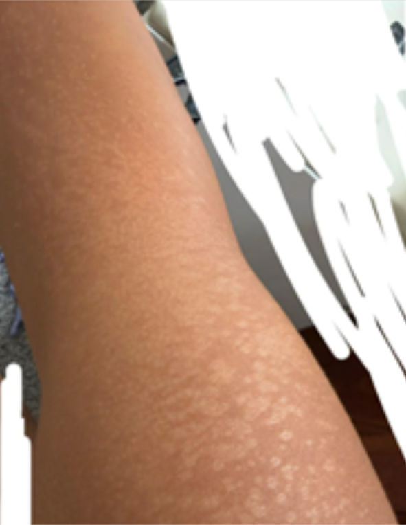
Quick Answer: Numerous small, fairly uniform, pale (hypopigmented) spots on the trunk without obvious fine scaling lean toward Progressive Macular Hypomelanosis (PMH) rather than classic pityriasis versicolor — though history is crucial: patches worsening in summer with fine scale on scratching favour versicolor, and prior eczema keeps post-inflammatory hypopigmentation in the frame. PMH is not contagious and not caused by poor hygiene. Treatment options include antibacterial washes such as benzoyl peroxide, topical antibiotics, and controlled sunlight or phototherapy in selected cases — with gradual improvement over months. Case Snapshot: A patient described white spots and patches slowly growing bigger on the trunk. The images showed numerous small, fairly uniform hypopigmented macules without the obvious fine branny scaling typical of pityriasis versicolor — tipping the assessment, cautiously, toward PMH, with the differentials explicitly kept open. The Three-Way Differential Pale spots on a young adult's trunk have three regular explanations, and telling them apart changes treatment entirely: 1. Progressive Macular Hypomelanosis (PMH) — the front-runner here: numerous small, fairly uniform hypopigmented macules, typically on the trunk (especially the back and lower back), classically without prominent scale, often slowly coalescing. PMH is linked to a variant of Cutibacterium bacteria in follicles subtly dialling down local pigment production. 2. Pityriasis versicolor — the Malassezia yeast overgrowth: the crucial distinguishers being worsening during summers/sweating and fine branny scale demonstrable on scratching the patches. If those features are present, versicolor remains an important differential — and it treats with antifungals, which do nothing for PMH. 3. Post-inflammatory hypopigmentation — pale patches at sites of previous inflammation: since this patient gives a history of eczema, it remains a close differential, typically showing patches that map onto where the eczema was. Why the Distinction Matters in Practice These three conditions are serially mistaken for each other — and the classic patient journey is months of antifungal creams for what was actually PMH, with predictable non-response and mounting worry. The bedside separators are simple in trained hands: the scratch test for scale, the distribution pattern, Wood's lamp examination (PMH follicles show characteristic dots of fluorescence; versicolor glows differently), and where needed a KOH scraping to confirm or exclude yeast in minutes. The Reassurances PMH Patients Need Two sentences do a lot of therapeutic work here: PMH is not contagious and is not caused by poor hygiene. It is also not vitiligo — the spots are hypopigmented (reduced pigment), not the stark depigmented white of vitiligo — and not dangerous. What it is: a slow, benign, cosmetically annoying pigment dimming that responds to correctly targeted treatment. Treatment — Aimed at the Actual Cause Because PMH is bacteria-associated, treatment logically targets bacteria, not yeast: Antibacterial washes such as benzoyl peroxide over the affected zones Topical antibiotics as prescribed Controlled sunlight or phototherapy in selected cases — stimulating the underactive pigment cells to switch back on And the expectation line that prevents premature abandonment: improvement is often gradual and may take several months — pigment recovery is intrinsically slow, so the plan is judged in quarters, not weeks. Photograph monthly; the camera sees what the mirror can't. Tracking Progress Without Losing Heart Because pigment recovery is measured in months, structure the follow-up: photograph a fixed reference area monthly under the same light; expect the earliest change as softening contrast rather than disappearance; review with your dermatologist at the three-month mark to confirm the trajectory or adjust; and hold treatment consistency through the unimpressive middle weeks — the phase where most patients quit a plan that was actually working. Sun sense throughout (the surrounding skin tanning less keeps the spots less visible) quietly does as much for appearance as the treatment itself. When to See a Dermatologist A dermatology evaluation may be needed to confirm the diagnosis and choose the most appropriate treatment — this case is the textbook argument for it, since three look-alike conditions with three different treatments are on the table, separable in one visit with a lamp and a scraping. Also return if patches turn stark white, sharply bordered, numb, or scaly-and-spreading — the cues to re-examine. Book a pigmentation assessment here, and explore our broader pigmentation services. FAQs Q: Is this early vitiligo? PMH spots are pale, not paper-white, with soft borders and a trunk-clustered pattern — quite different from vitiligo under examination and Wood's lamp. The visit that names PMH also credibly excludes vitiligo. Q: I used antifungal creams for months with no change — why? Very likely because the diagnosis was PMH (or post-eczema hypopigmentation), which antifungals don't touch. Non-response to a correct-seeming treatment is itself a diagnostic clue. Q: Will the pale spots repigment fully? PMH typically improves substantially with targeted treatment and time, though slowly; some cases also settle spontaneously over years. Q: Can I speed things up by sunbathing? Unstructured sun deepens the surrounding tan and can worsen contrast. Light-based treatment works — as controlled phototherapy, dosed by a dermatologist. 📌 Case Study Reference: Publicly shared patient images and query, r/DermatologyQuestions, Reddit — "White spots and patches slowly growing bigger on…". Educational discussion only; not a final diagnosis. Clinical commentary by the Allodermis dermatology team.

Learn more

AI Skin Analysis