







Skip to content
Up to 30% Off on all Treatments

Case Study

Case 14: Forearms Darker Than the Rest of Your Body? Phototanning and Uneven Skin Tone Explained

Case 14: Forearms Darker Than the Rest of Your Body? Phototanning and Uneven Skin Tone Explained

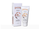
Quick Answer: When sun-exposed skin (forearms, lower arms, face) becomes visibly darker than covered skin, with a clear demarcation line at the sleeve, the diagnosis is usually phototanning — UV-stimulated melanin production. The most important treatment is strict photoprotection: full sleeves or UV arm sleeves plus broad-spectrum sunscreen reapplied outdoors. Tan reduction is then achieved gradually with glycolic, lactic, or kojic acid, with dermatologist-supervised body peels for faster results. Case Snapshot: An Indian male presented with darkening of the sun-exposed skin of his forearms and lower arms, with a sharp line of demarcation where his half-sleeve shirt ended — the classic "driver's tan" pattern. What Is Phototanning? Phototanning is the skin's natural defence at work: UV exposure stimulates melanocytes to produce more melanin, darkening the exposed skin. The result is a two-tone appearance — photoexposed areas conspicuously darker than photoprotected ones — with a clear demarcation line that patients often find more distressing than overall darkness would be. The Everyday Habits Behind It Driving with exposed arms — window-side arms tan asymmetrically Outdoor sports and commuting through peak sun hours Half-sleeve clothing creating the sharp sleeve line Inconsistent sunscreen use — applied some days, forgotten most Because these exposures repeat daily, the tan deepens continuously; treatment without behaviour change is a bucket with a hole in it. The Treatment Ladder Strict photoprotection — the foundation. Full-sleeve clothing or UV-protective arm sleeves, plus broad-spectrum sunscreen on all exposed skin, reapplied every 2–3 hours outdoors. No de-tanning plan works without this step. Topical tan reduction: body lotions with glycolic acid, lactic acid, or kojic acid gently accelerate pigment turnover over weeks. Dermatologist-supervised body peels for faster, more uniform results — a series of controlled chemical peels designed for body skin. Oral photoprotective supplements — useful as an adjunct only, never a replacement for sunscreen. Clinic-based de-tan programs: explore our dedicated tanned skin treatments and face & body tan removal services. Realistic Timelines A tan is living biology, not a stain. With airtight photoprotection, superficial tanning fades noticeably over 6–12 weeks as pigmented cells turn over; acids and peels shorten this. The demarcation line softens last. Patients who continue daily unprotected exposure, however, will out-tan any treatment — which is why we treat the habit first and the pigment second. When a "Tan" Is Not a Tan Persistent or patchy darkening on exposed skin can occasionally represent other photo-aggravated conditions — certain drug-induced pigmentations, melasma extending onto the arms, or dermatitis-related pigment. Clues that warrant evaluation: itching, scaling, patchy rather than uniform darkening, or pigmentation that ignores the sun-exposure pattern. Our hyperpigmentation team can differentiate these on examination. When to See a Dermatologist If tanning is persistent despite two months of genuine photoprotection, if pigmentation is uneven or symptomatic, or if you want a supervised body-peel program before an event, book a consultation. FAQs Q: Which SPF should I use for Indian sun? Broad-spectrum SPF 50 with PA+++ or higher, applied generously and reapplied outdoors. Quantity and reapplication matter more than the number on the tube. Q: Do de-tan face packs and home remedies work? Mild exfoliating effects at best; lemon and similar acidic remedies often irritate and worsen pigmentation. Structured acids plus sunscreen outperform them safely. Q: Will my original skin tone come back completely? Usually yes for recent tanning, over weeks to months of protection. Years of cumulative exposure may leave residual pigment that needs peels or laser-assisted treatment. Q: Are glutathione drips a shortcut? They are not a substitute for photoprotection, and should only ever be considered after a dermatologist consultation about evidence, safety, and expectations. Photoprotection That Survives Real Indian Life Sunscreen advice fails when it ignores logistics, so here is the field-tested version. Quantity: roughly half a teaspoon for the face and neck, and a full teaspoon per forearm — most people apply a quarter of this. Timing: apply 15 minutes before leaving; keep a small tube in your bag or vehicle for the 2–3-hourly reapplication that outdoor days demand. For two-wheeler riders and drivers — the classic phototanning population — UV arm sleeves are transformative: they outperform sunscreen for arms because they cannot wear off, sweat off, or be forgotten, and they cost less than a month of sunscreen. Peak hours: where schedules allow, shift outdoor errands away from 11 am–3 pm. Don't forget the neglected zones that betray a de-tan effort: the back of the neck, the ears, the V of the chest, and the backs of the hands. Once this armour is in place, every rupee spent on de-tan actives and peels finally stays spent — the pigment you remove no longer gets replaced by tomorrow's commute. 📌 Case Study Reference: Publicly shared patient image and query, Reddit — "Solutions to this?" (forearm tanning image post). Educational discussion only; not a final diagnosis. Clinical commentary by the Allodermis dermatology team.

Learn more
Case 13: "Retinol Is Making My Skin Worse" — Retinoid Dermatitis and the First-Month Purge Explained

Case 13: "Retinol Is Making My Skin Worse" — Retinoid Dermatitis and the First-Month Purge Explained

Quick Answer: Redness, peeling, and a flare-up of acne in the first month of retinoid use is common and usually temporary. It happens because the skin's surface initially over-responds to retinoids and because deep-seated acne is pushed to the surface as follicles loosen. It is managed by temporarily reducing the frequency, amount, or duration of application and supporting the skin with moisturisers — not by quitting. Uncontrolled or severe reactions need a dermatologist's review. Case Snapshot: An Indian female patient complained of significant redness and a sudden flare of acne after starting a retinoid, sharing photos captioned "retinol is doing shit" — a frustration dermatologists hear weekly, and one with a clear biological explanation. Why Retinoids Get Worse Before Better Two separate processes drive the rocky first month: Retinoid dermatitis — a hyperproliferative response of the epidermis: the surface skin initially turns over too fast, producing visible redness, dryness, and peeling before it adapts. The purge — retinoids loosen the follicular lining, causing externalisation of deep-seated acne: microcomedones that were already forming beneath the surface erupt earlier than they otherwise would. This looks like the retinoid "causing" acne; in reality it is fast-forwarding lesions that were coming anyway. Both phenomena peak in the first 2–6 weeks and settle as the skin retinises (adapts). How Dermatologists Bring It Under Control Temporarily reduce the frequency — from nightly to alternate nights or twice weekly Reduce the amount — a pea-sized quantity covers the whole face; more is not better Reduce the duration — short-contact application (apply for 30–60 minutes, then wash off) allows sensitive skin to adapt gradually Moisturise generously — before and after the retinoid ("sandwiching") buffers irritation without meaningfully reducing efficacy Simplify everything else — pause acids, scrubs, and other actives while the skin adapts; keep only a gentle cleanser, moisturiser, and morning sunscreen Once tolerance builds, frequency and strength are stepped back up — the destination is unchanged, only the pace changes. Purge vs True Reaction: How to Tell A purge occurs in your usual acne-prone zones, consists of typical pimples, and steadily improves after weeks. A true irritant or allergic reaction brings burning, intense itching, swelling, or rash in new areas — including places you never break out. This warrants stopping and seeing a dermatologist promptly, sometimes with patch testing to identify the culprit. What Not to Do Do not scrub the peeling skin, do not layer multiple new products to "fix" the irritation, and do not abandon retinoids permanently after one rough fortnight — they remain the most evidence-backed molecules in dermatology for acne, texture, and ageing. Equally, do not push through severe, uncontrolled reactions out of stubbornness; escalating redness and pain is a medical signal, not a rite of passage. When to See a Dermatologist Visit a dermatologist if irritation is severe, spreading, or not settling with reduced frequency and moisturiser support — or if the "purge" continues beyond 8–10 weeks, which suggests the regimen needs restructuring. Supervised titration (as in our clinic protocols) gets patients through adaptation with far less misery. Book a review here, and read our full guide to acne treatment timelines. FAQs Q: How long does the retinoid purge last? Typically 2–6 weeks, occasionally up to 8. Beyond that, the regimen should be reviewed. Q: Should I stop retinol completely if my face peels? Usually no — reduce frequency and buffer with moisturiser. Stop and seek review only for severe burning, swelling, or rash. Q: Can I use retinoids in Indian summers? Yes — retinoids are night products. The non-negotiable partner is diligent morning sunscreen, season regardless. Q: Is prescription tretinoin harsher than cosmetic retinol? Generally yes, and also more effective. Dermatologist-guided titration makes even tretinoin tolerable for most skin. A Week-by-Week Map of Retinisation Knowing what "normal" looks like keeps patients on course: Weeks 1–2: mild tightness, dryness, and fine flaking; possibly the first purge pimples in usual breakout zones Weeks 3–5: the trough — peak purging and peak irritation for many; this is where most people wrongly quit Weeks 6–8: irritation subsides; purging thins out; early smoothness appears Weeks 9–12: visibly clearer, more even skin; frequency and strength can now be advanced under guidance Months 4–6+: the compounding phase — texture, marks, and pore appearance keep improving If your trajectory diverges sharply from this map — worsening after week 8, burning that intensifies rather than fades, or rash in non-acne zones — that is the signal to book a review rather than push through. And two practical safeguards throughout: apply retinoids to fully dry skin (damp skin absorbs more and stings more), and keep the product away from the eyelids, nostril creases, and lip corners, where irritation concentrates first. 📌 Case Study Reference: Publicly shared patient images and query, Reddit — "Retinol is doing shit" (image post). Educational discussion only; not a final diagnosis. Clinical commentary by the Allodermis dermatology team.

Learn more
Case 12: Dark Spots Left Behind After Acne — Post-Inflammatory Hyperpigmentation (PIH) in Indian Skin

Case 12: Dark Spots Left Behind After Acne — Post-Inflammatory Hyperpigmentation (PIH) in Indian Skin

Quick Answer: Dark spots after acne are post-inflammatory hyperpigmentation (PIH) — the skin's response to inflammation, producing extra melanin at the exact spot where a pimple healed. PIH is especially common in Indian and darker skin tones. There is no overnight fix, but it is very treatable with lightening agents such as azelaic acid, arbutin, kojic acid, and glycolic acid, plus chemical peels — and the best strategy is prevention: treat acne early, never pick, and protect from the sun. Case Snapshot: A user on r/IndianSkincareAddicts shared photos asking for help with skin texture and "black marks" scattered across the face — flat brown spots precisely marking where previous pimples had been. Why Acne Leaves Marks on Indian Skin When a pimple inflames, the skin responds to that inflammation by producing extra melanin (pigment) at the same spot. In richly pigmented skin — most Indian skin tones — melanocytes are more reactive, so even a modest pimple can leave a stubborn brown mark. Worse, when inflammation is intense or the lesion is squeezed, pigment gets deposited deeper in the skin, where it becomes far harder to clear. That is why the marks often outlast the acne itself by months. Mark or Scar? Get This Right First PIH (mark): flat, brown or dark, smooth to touch — a colour problem. Fades with time and treatment. Scar: a texture change — pit, depression, or unevenness — a collagen problem. Will not fade with creams; needs procedural scar treatment. Many faces carry both, and each needs its own plan. Prevention: The Three Rules Treat acne early — every inflamed pimple that never happens is a mark that never forms. Start proper acne treatment at the first sign of recurring breakouts. Never pick or squeeze — manipulation multiplies inflammation and pushes pigment deeper. Protect from the sun daily — UV exposure darkens existing marks and dramatically slows their fading. Broad-spectrum sunscreen every morning is the single most powerful anti-PIH habit. Treatment: No Quick Fix, But Real Results Azelaic acid — the workhorse for PIH in Indian skin: it lightens marks and treats acne simultaneously, with an excellent safety profile Arbutin and kojic acid — melanin-pathway inhibitors used in dermatologist-guided combinations Glycolic acid — accelerates pigment turnover through gentle exfoliation Chemical peels in-clinic — speed up clearance of superficial pigment over a series of sessions Laser toning for deeper, resistant pigment, at conservative settings appropriate for darker skin Expect gradual fading over 3–6 months for superficial marks; deeper pigment takes longer. Consistency plus sunscreen beats any single "miracle" product. The Trap to Avoid Steroid-containing "fairness" combination creams lighten marks briefly, then cause rebound darkening, thinning, and acne flares of their own. They are the most common way patients convert a solvable pigment problem into a chronic one. Use only dermatologist-recommended actives — our skin pigmentation page explains the safe options. When to See a Dermatologist If marks are widespread, deepening, or not fading after three months of sunscreen and actives — or if active acne is still producing new marks — see a dermatologist. Controlling the acne and clearing the pigment together shortens the whole journey. Book a consultation. FAQs Q: How long do acne marks take to fade naturally? Superficial PIH fades over 3–6 months with sun protection; deeper pigment can take a year or more without treatment — actives and peels significantly shorten this. Q: Does vitamin C help PIH? Yes — as an antioxidant brightener it supports the plan, though azelaic acid and peels do the heavier lifting for established marks. Q: Will the marks come back after treatment? Not the same ones — but new acne creates new marks. That's why acne control and maintenance are part of every PIH plan. Q: Are red marks and brown marks treated the same way? Red marks (post-inflammatory erythema) are residual blood-vessel changes and fade with time or vascular-targeted treatment; brown marks are melanin and respond to the lightening agents above. Many lesions carry both, fading red-first, brown-second. A Daily Anti-PIH Routine Morning: gentle cleanser → azelaic acid (if prescribed for daytime) → moisturiser → broad-spectrum SPF 50, reapplied outdoors Night: cleanser → retinoid or lightening active as guided → moisturiser Weekly discipline: hands off the face; no picking, no "checking" bumps in the mirror with fingernails Monthly: photograph the marks in identical lighting — fading is too slow to see daily but obvious month-to-month, and the visible progress protects motivation How Clinic Treatments Are Sequenced In-clinic care follows a logic: first stabilise acne (so the mark factory closes), then superficial peels every few weeks to accelerate pigment turnover, then laser toning only for the stubborn deeper remainder. Jumping straight to procedures while acne still rages wastes sessions — each new pimple deposits fresh pigment behind the treatment. This sequencing conversation is exactly what a first consultation establishes. 📌 Case Study Reference: Publicly shared patient images and query, r/IndianSkincareAddicts, Reddit — Want to fix skin texture / black marks (image post). Educational discussion only; not a final diagnosis. Clinical commentary by the Allodermis dermatology team.

Learn more
Case 11: Painful Thick Skin on the Sole — Is It a Corn or a Plantar Wart? Most People Treat the Wrong Thing

Case 11: Painful Thick Skin on the Sole — Is It a Corn or a Plantar Wart? Most People Treat the Wrong Thing

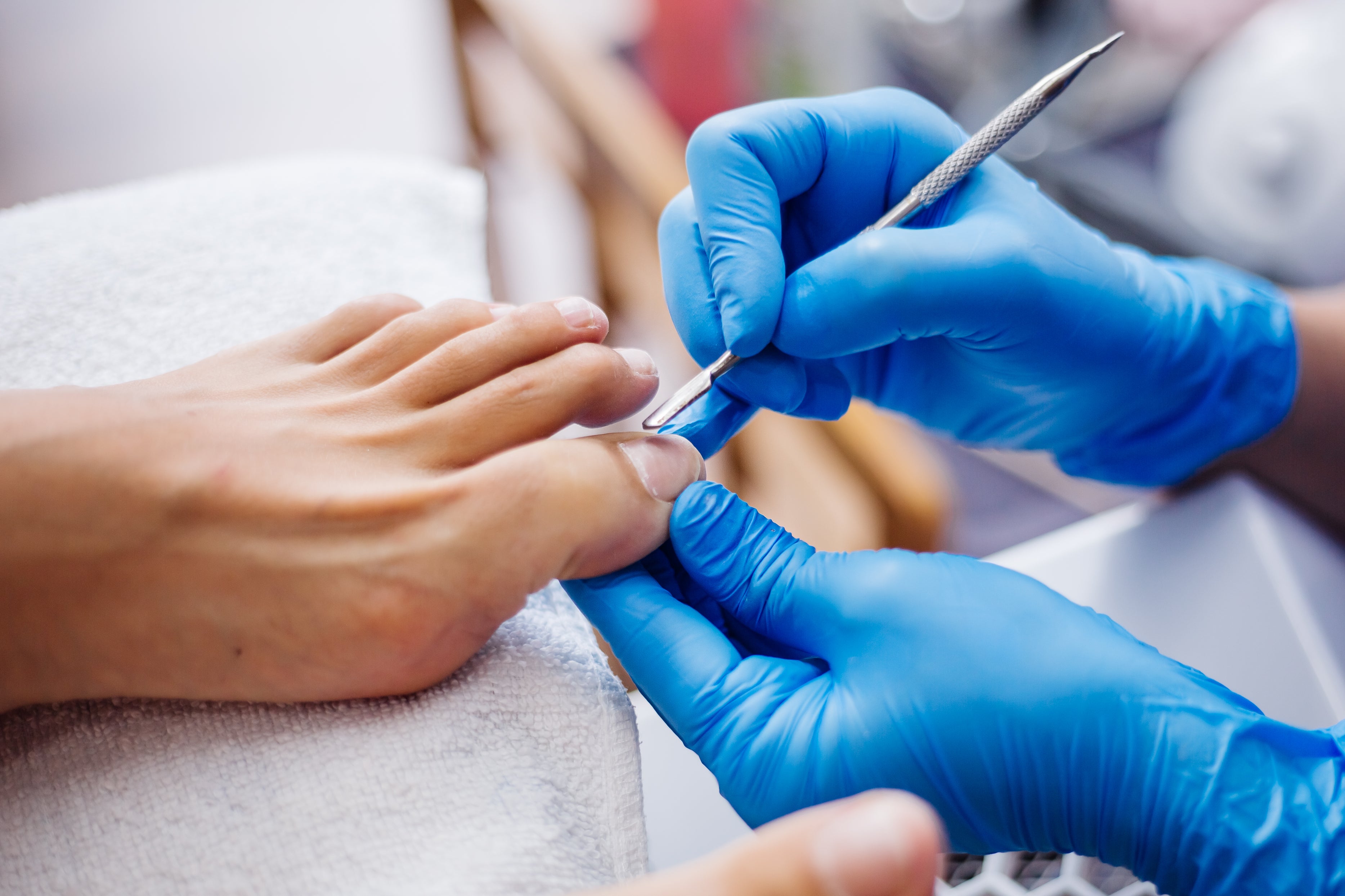
Quick Answer: A corn is caused by pressure and friction, forms a hard central core, and hurts on direct downward pressure. A plantar wart is caused by HPV infection, shows tiny black or red dots (clotted blood vessels), interrupts the normal skin lines, and typically hurts more on side-to-side squeezing. Corns need pressure relief and keratolytics; warts need salicylic acid, cryotherapy, or dermatologist-supervised procedures. Treating the wrong one worsens the pain. Case Snapshot: A patient presented with a painful, thickened patch on the sole of the foot that they had been shaving down at home for months, assuming it was a corn. Close examination revealed pinpoint black dots and interrupted skin lines — the fingerprint-like ridges of the sole stopped at the lesion's edge — pointing instead to a plantar wart. Corn vs Wart: The Diagnostic Clues That Matter Corn: a response to chronic pressure and friction — typically over bony points. It has a hard central core, preserved skin lines running through it, and is painful on direct downward pressure (stepping on it). Plantar wart: a viral infection with HPV (human papillomavirus). Its tell-tale signs are tiny black or red dots — clotted capillaries, highly typical of warts — interrupted skin lines, and greater pain on side-to-side squeezing than on direct pressure. These small differences completely change management, which is why self-diagnosis so often fails on the foot. How Plantar Warts Are Caught HPV enters through microscopic breaks in the skin. Risk rises with: Walking barefoot in wet public places — gyms, swimming pools, changing rooms Minor skin trauma on the sole Excessive sweating, which softens and macerates the skin Lowered immunity Treatment: Two Different Roads For corns: the cause is mechanical, so the cure is mechanical — pressure relief with proper footwear, protective padding, and keratolytics to thin the hard skin. Without addressing the footwear and pressure point, corns always return. For plantar warts: the cause is viral, so treatment must destroy infected tissue — salicylic acid preparations, cryotherapy (liquid nitrogen), electrocautery, laser, or needling, all under dermatologist supervision. Multiple sessions are often needed, and stubborn warts sometimes require immunotherapy approaches. Explore professional wart removal at Allodermis for safe, definitive treatment. Why Home Treatment Backfires Picking or shaving a plantar wart at home does two dangerous things: it spreads the virus to surrounding skin (creating satellite warts) and drives the lesion deeper, making it more painful and harder to treat. Conversely, freezing or burning a corn as if it were a wart injures healthy pressure-bearing skin without removing the mechanical cause. In both directions, treating the wrong lesion worsens pain — the central lesson of this case. The Footwear Factor Whether corn or wart, the sole's environment matters. Well-fitted, cushioned footwear reduces the pressure that creates corns and the micro-trauma that admits HPV. Moisture-wicking socks and drying the feet thoroughly reduce the softened-skin conditions warts favour. Patients who fix their footwear alongside treatment relapse far less. When to See a Dermatologist See a dermatologist if a sole lesion is painful, growing, bleeding, multiplying, or has resisted a month of over-the-counter care — or simply if you are unsure which lesion you have. Diabetic patients should never self-treat foot lesions at all; even minor procedures on diabetic feet require medical supervision. Book an evaluation here. FAQs Q: Are plantar warts contagious to family members? Yes — through shared floors, bathrooms, and footwear. Keep feet covered at home during treatment and avoid sharing slippers and towels. Q: How long does wart treatment take? Salicylic acid regimens run weeks to months; cryotherapy and procedural options typically need multiple sessions spaced 2–3 weeks apart. Persistence wins. Q: Can a wart go away on its own? Some do, over months to years, as immunity catches up — but on the weight-bearing sole they usually deepen and spread first, so treatment is recommended. Q: Why does my "corn" keep coming back after removal? Either the pressure source (footwear, bony prominence) was never corrected — or it was never a corn. Recurrence is a classic sign of a misdiagnosed plantar wart. Preventing the Next Foot Lesion A few habits protect against both problems long-term: wear waterproof slippers in gyms, pool decks, and shared changing rooms — the classic HPV transmission zones; dry the feet thoroughly, especially between the toes, and change sweaty socks promptly; inspect the soles monthly so lesions are caught small, when treatment is quickest; replace worn footwear whose collapsed cushioning creates new pressure points; and never share nail clippers, pumice stones, or foot files, which can carry the virus between people and between sites on your own feet. If you've had one plantar wart, be extra vigilant — a previously infected sole plus the same barefoot habits is how "one wart" becomes a mosaic of many. And a final word on the popular home approach of paring lesions with blades: beyond spreading virus, it risks wounds and infection, and in anyone with diabetes or reduced foot sensation it is genuinely dangerous. Professional wart removal is faster, safer, and — because the diagnosis is confirmed first — actually aimed at the right target. 📌 Case Study Reference: Publicly shared patient image and query, Reddit dermatology/skincare communities. Educational discussion only; not a final diagnosis. Clinical commentary by the Allodermis dermatology team.

Learn more
Case 10: Sandpaper-Like Bumps on Arms and Thighs — Keratosis Pilaris ('Chicken Skin') Explained

Case 10: Sandpaper-Like Bumps on Arms and Thighs — Keratosis Pilaris ('Chicken Skin') Explained

Quick Answer: Tiny rough bumps on the arms or thighs that feel like sandpaper are usually keratosis pilaris (KP) — a very common, harmless, hereditary condition where keratin (dead skin protein) plugs the hair follicles. The most effective treatment is deep, consistent moisturisation using creams with urea, lactic acid, glycolic acid, or salicylic acid. Hard scrubbing makes it worse, and results take time — consistency is key. Case Snapshot: A patient described persistent rough, skin-coloured to reddish bumps over the upper arms that felt like sandpaper. They had been scrubbing the area vigorously for months, only to find the bumps looking more inflamed and prominent. What Is Keratosis Pilaris? Keratosis pilaris occurs when dead skin protein (keratin) blocks the openings of hair follicles, producing hundreds of tiny, rough, skin-coloured or reddish bumps — classically on the upper arms and thighs, sometimes the cheeks and buttocks. It is: Extremely common — one of the most frequent skin findings worldwide Completely harmless — a cosmetic texture issue, not a disease or infection Often familial — it runs strongly in families, reflecting an inherited keratinisation tendency Why It Flares Dry skin — KP and dryness feed each other Winter weather and low humidity Harsh soaps that strip the barrier Friction from clothing The Treatment That Actually Works The strategy is chemical softening, not physical force: Deep moisturisation as the foundation — twice daily, every day Keratolytic creams containing urea, lactic acid, glycolic acid, or salicylic acid — these gently dissolve the keratin plugs and smooth the surface over weeks Gentle cleansing with soap-free washes; lukewarm showers Consistency over intensity — results build gradually over 4–8 weeks and are maintained only with continued care The Mistake Almost Everyone Makes Scrubbing too hard. Physical scrubs and loofahs feel productive but actually worsen dryness, inflame the follicles, and make the bumps more visible and red. KP is a keratin chemistry problem; it responds to acids and hydration, not abrasion. Setting Expectations KP is a chronic but very manageable condition. It typically improves through the 20s and 30s and fluctuates with seasons. There is no permanent cure, but with the right routine, skin can stay consistently smooth. When redness and dotted pigmentation persist, in-clinic options — prescription-strength keratolytics, gentle chemical peels, and in select cases laser toning for associated pigmentation — can accelerate and refine results. When to See a Dermatologist Consult a dermatologist if redness, itching, or cosmetic concern persists despite 6–8 weeks of proper moisturisation and keratolytics, or if you're unsure whether your bumps are truly KP — folliculitis, eczema, and other follicular conditions can mimic it and need different treatment. Book a consultation with our team for a personalised body-care plan. FAQs Q: Does keratosis pilaris go away on its own? It often softens with age and improves in humid weather, but it tends to flare in winter. Maintenance moisturisation keeps it controlled year-round. Q: Is chicken skin contagious? No — KP is an inherited keratinisation tendency, not an infection. It cannot spread to others or to other body parts by touch. Q: Which strength of actives should I use? Typical effective ranges are urea 10%+, lactic acid ~12%, glycolic acid 6–12%, or salicylic acid 2%+. A dermatologist can prescribe stronger formulations if over-the-counter options plateau. Q: Why do my KP bumps look red or leave dark dots? Redness reflects follicular inflammation (keratosis pilaris rubra pattern); the dark dots are post-inflammatory pigmentation around each follicle — the "strawberry skin" appearance. Both respond to the same anti-KP routine, with the pigmentation fading gradually once inflammation and friction stop. A Simple KP Body Routine Shower: lukewarm, under 10 minutes, soap-free wash; no loofah on affected areas Immediately after: keratolytic lotion (urea/lactic/glycolic/salicylic) over the bumps, plain moisturiser everywhere else Night: repeat the keratolytic on the worst zones; layer a thicker cream over it in winter Weekly: at most, a very gentle chemical exfoliation — never physical scrubbing Clothing: soft, breathable fabrics; avoid tight sleeves that rub the upper arms KP and "Strawberry Skin" Before Events Many patients seek fast fixes before weddings or holidays. The honest guidance: start the routine at least 6–8 weeks ahead — that's how long keratolytics need to visibly smooth texture and calm redness. For residual dotted pigmentation on the arms, dermatologist-supervised options such as gentle body chemical peels can add polish, and where coarse hair contributes to the bumpy look, laser hair reduction reduces both the texture and the ingrown-hair component. Last-minute aggressive scrubbing, by contrast, guarantees redder, angrier arms on the day itself. 📌 Case Study Reference: Publicly shared patient images and queries, Reddit skincare communities (see related query: Chicken skin on my arms and shoulders — product help). Educational discussion only; not a final diagnosis. Clinical commentary by the Allodermis dermatology team.

Learn more
Case 9: Acne Scars CAN Be Prevented — and Treated. Here's the Full Picture

Case 9: Acne Scars CAN Be Prevented — and Treated. Here's the Full Picture

Quick Answer: Acne scars form when deep inflammation destroys the skin's collagen, leaving pits, depressions, and uneven texture — most often on the cheeks. The single biggest preventable cause of worsening scars is picking or squeezing pimples. Early acne treatment under dermatologist guidance is the best prevention; existing scars improve significantly with microneedling, subcision, TCA CROSS, and laser treatments. Case Snapshot: A user on r/IndianSkincareAddicts shared images of pitted cheek scarring left behind by past breakouts and asked for product suggestions — highlighting a common misconception that scars can be fixed with skincare products alone. How Acne Scars Actually Form When acne inflammation penetrates deep into the dermis, it damages the skin's structural collagen. As the skin heals, it either produces too little collagen (creating atrophic scars — pits and depressions) or occasionally too much (raised hypertrophic scars). The cheeks are the most common battleground, and once collagen architecture is lost, no cream can rebuild it — which is why prevention is everything. The #1 Cause of Worsening Scars Picking and squeezing pimples. Squeezing multiplies inflammation and pushes infected material deeper into the dermis, converting a lesion that might have healed cleanly into one that scars permanently. If one habit change protects your future skin, it is this: hands off. The Prevention Playbook Start acne treatment early — every month of untreated inflammatory acne adds scar risk Use dermatologist-guided medications: retinoids, benzoyl peroxide, or prescription therapy matched to your severity Control breakouts fully rather than partially — smouldering low-grade acne still scars Never pick — get painful lesions treated in-clinic instead For the complete acne roadmap, read Acne Treatment Explained, and explore our pimple treatment services. Treating Scars That Already Exist Modern dermatology offers genuinely effective, collagen-remodelling procedures — usually in combination: Microneedling — controlled micro-injury that stimulates fresh collagen MNRF (microneedling radiofrequency) — adds radiofrequency energy at depth for rolling and boxcar scars Subcision — releases fibrous bands tethering rolling scars downward TCA CROSS — high-strength acid applied precisely into deep ice-pick scars Fractional laser resurfacing — refines overall texture Because most patients have a mix of scar types, combination protocols tailored to each scar deliver the best outcomes. See our full acne scar treatment options and the detailed guide Acne Scar Treatment Explained — Types, Best Treatments and Timelines. Realistic Expectations Complete scar erasure is uncommon; meaningful, visible improvement of 50–80% is achievable over 6–12 months with 3–8 sessions, depending on scar type and depth. Improvement continues for months after each session as collagen matures. Consistency and the right combination matter more than any single "miracle" procedure. When to See a Dermatologist If you currently have active acne, see a dermatologist now — that is scar prevention in real time. If you have established scars, book an assessment so each scar type can be mapped and matched to the right procedure. Schedule your consultation here. FAQs Q: Can acne scars disappear completely? Complete removal is uncommon, but combination therapy typically achieves 50–80% visible improvement over 6–12 months. Q: How many sessions will I need? Usually 3–8 sessions, spaced weeks apart, depending on scar type, depth, and the procedures combined. Q: Do scar creams work on pitted scars? No cream rebuilds lost collagen at depth. Creams help surface marks (pigmentation), while pitted scars need procedural treatment. Q: What's the difference between ice-pick, boxcar, and rolling scars? Ice-pick scars are narrow, deep pits; boxcar scars are wider depressions with sharp edges; rolling scars are shallow waves tethered from below. Each responds best to a different procedure — TCA CROSS, resurfacing, and subcision respectively — which is why scar mapping precedes treatment. The Numbers Behind Acne Scarring Scarring is not a rare complication — it is the expected outcome of unmanaged inflammatory acne. Studies suggest that up to 90% of acne patients develop some degree of scarring, and roughly 22% suffer significant scarring. The variables that tip a patient into the severe group are consistent: delayed treatment, deeper inflammatory lesions (nodules and cysts), picking, and genetic healing tendencies. The practical implication is powerful — the timing of your first dermatology visit may matter more for your future skin than any procedure done later. Aftercare That Protects Your Investment Scar procedures create controlled injury; how you heal determines the payoff: Strict sunscreen for weeks after each session — new collagen and healing skin pigment easily in Indian skin tones No picking at treated skin, crusts, or peeling Gentle skincare only for the first days: bland cleanser and moisturiser, resuming retinoids on your dermatologist's schedule Space sessions correctly — collagen remodelling takes 4–6 weeks between treatments; rushing adds risk, not speed Photograph monthly in identical lighting to track genuine progress 📌 Case Study Reference: Publicly shared patient images and query, r/IndianSkincareAddicts, Reddit — https://www.reddit.com/r/IndianSkincareAddicts/comments/1shy5p1/product_suggestions_plzzz/. Educational discussion only; not a final diagnosis. Clinical commentary by the Allodermis dermatology team.

Learn more
Case 8: Tiny White-Yellow Bumps on the Lips — Fordyce Spots Are Normal, Not an STD

Case 8: Tiny White-Yellow Bumps on the Lips — Fordyce Spots Are Normal, Not an STD

Quick Answer: Painless tiny white or yellow bumps on the lips are usually Fordyce spots — naturally occurring, visible oil (sebaceous) glands. They are completely harmless, not infectious, and not a sexually transmitted disease. No treatment is required, though cosmetic options like topical retinoids or laser exist for those who are bothered by their appearance. Case Snapshot: An anxious patient asked about small white-yellow bumps along the border of the lips that neither hurt nor itched, worried they might be an infection or something sexually transmitted. What Are Fordyce Spots? Fordyce spots are normal sebaceous (oil) glands that happen to be visible on the lips or genital skin. Every human has sebaceous glands; in most areas they open into hair follicles and stay hidden, but on the lip border and genital skin they open directly onto the surface, where they can appear as tiny pale bumps. They are: ✅ Harmless — a normal anatomical variation, present in a large proportion of adults ✅ Not infectious — they cannot spread to another person ✅ Not a sexually transmitted disease — despite frequently causing STD anxiety Why Do They Suddenly Become Noticeable? Patients often report that the spots "appeared recently." In reality, the glands were always there and became more visible due to: Skin stretching (which spreads the overlying skin thinner) Dryness of the lips, reducing surface glow that camouflages them Hormonal changes that increase oil-gland activity, enlarging the glands slightly Do Fordyce Spots Need Treatment? Usually not. Dermatologists worldwide consider them a normal variant requiring only reassurance. If they are genuinely cosmetically bothersome, in-clinic options exist: Topical retinoids to modestly reduce prominence over time CO₂ laser, chemical cautery, electrocautery, or micro-punch excision in selected cases It is important to set expectations: results vary, and recurrence can occur, because the underlying glands are a permanent, normal part of the skin. Aggressive removal attempts risk scarring far more noticeable than the spots themselves. The Important Caveat: Not Every Lip Bump Is Fordyce Several conditions can mimic Fordyce spots — milia, flat warts, molluscum contagiosum, and occasionally early mucosal lesions. The distinguishing features (uniformity, distribution, absence of symptoms) are best judged in person, sometimes with dermoscopy. Getting a confirmed diagnosis before considering any procedure is essential; our skin diagnostic services and dermatologist consultations are designed exactly for this. The Psychological Side A large part of managing Fordyce spots is managing the anxiety around them. Patients frequently avoid relationships or feel embarrassed over what is, medically, as normal as a freckle. Clear information is the treatment: these are your own oil glands, everyone has them, and nothing is wrong. When to See a Dermatologist See a dermatologist if bumps change, grow, become painful, itch, ulcerate, or bleed — those features suggest a different diagnosis. Also consult before undergoing any cosmetic removal so a qualified doctor can confirm the diagnosis and discuss realistic outcomes. Book an appointment with our team here. FAQs Q: Can Fordyce spots spread to my partner? No — they are your own oil glands, not an infection, and cannot be transmitted. Q: Do lip balms remove Fordyce spots? No, but keeping lips well hydrated can make them appear less prominent. Q: Will Fordyce spots go away on their own? They typically persist as a normal skin feature, though prominence fluctuates with hydration and hormones. Q: Should I try to extract or pop Fordyce spots? Never. They are solid glands, not pimples — there is nothing to "pop." Squeezing only injures the lip border, risking marks, infection, and scarring in one of the most cosmetically visible zones of the face. How Dermatologists Confirm the Diagnosis Diagnosis is usually clinical: Fordyce spots present as multiple, uniform, 1–2 mm pale yellow-white bumps, symmetrically distributed along the vermilion border, painless and unchanging over time. Dermoscopy can help in doubtful cases, showing the characteristic yellowish lobular pattern of sebaceous glands. Features that argue against Fordyce spots — a single growing lesion, umbilicated (dimpled) bumps suggesting molluscum, rough warty surfaces, or any ulceration — prompt closer evaluation and occasionally a small skin biopsy for certainty. If You Do Choose Cosmetic Treatment For the minority who pursue removal, sensible ground rules apply: treat a test area first to preview results and healing; expect multiple sessions for laser-based approaches; protect healing skin with strict sun protection to prevent post-procedure pigmentation, especially in Indian skin; and accept that new glands can become visible later, since the treatment removes visible glands, not the tendency. Costs, downtime (usually a few days of mild redness or crusting), and realistic before-and-after expectations should all be discussed at consultation — a good dermatologist will talk as many patients out of treatment as into it, because reassurance genuinely is the best medicine here. 📌 Case Study Reference: Publicly shared patient image and query, r/IndianSkincareAddicts community, Reddit — https://www.reddit.com/r/IndianSkincareAddicts/. Educational discussion only; not a final diagnosis. Clinical commentary by the Allodermis dermatology team.

Learn more
Case 7: Dark Underarms — Friction, Shaving Damage, or a Metabolic Warning Sign?

Case 7: Dark Underarms — Friction, Shaving Damage, or a Metabolic Warning Sign?

Quick Answer: Dark underarms are most commonly caused by friction, repeated shaving or waxing irritation, dead skin buildup, and post-inflammatory pigmentation. However, thick, velvety, symmetrical darkening may be acanthosis nigricans — an early skin marker of insulin resistance, PCOS, or obesity that warrants medical screening. Treatment combines gentle AHA exfoliation, dermatologist-guided lightening creams, laser hair reduction, and management of any underlying metabolic cause. Case Snapshot: A patient sought help for persistent underarm darkening that had not responded to home remedies, natural masks, or over-the-counter whitening creams. She shaved regularly and wore fitted clothing daily. Why Underarms Darken The underarm is a high-friction, frequently traumatised skin fold. Common contributors include: Friction from clothing and skin-on-skin rubbing Repeated shaving or waxing — micro-injury after micro-injury triggers pigment Buildup of dead skin and residue from deodorants Post-inflammatory pigmentation from folliculitis, ingrown hair, or irritation Hormonal and metabolic factors The Warning Sign You Shouldn't Ignore When underarm skin becomes dark, thickened, and velvety — often symmetrically, and sometimes with similar changes on the neck — it may be acanthosis nigricans. This is not simple pigmentation; it is a skin signal of insulin resistance, commonly associated with PCOS and obesity, and can be an early warning of pre-diabetes. Such patients need screening (blood sugar, insulin, hormonal work-up) rather than just creams. Learn how hormones affect skin in our guide: PCOS Symptoms in Women — How Hormonal Imbalance Affects Skin and Hair. The Treatment Ladder Gentle chemical exfoliation with AHAs — glycolic or lactic acid lotions — to lift dead-cell buildup without friction Urea-based moisturisers to soften thickened skin Dermatologist-guided lightening creams: azelaic acid, kojic acid, or niacinamide Laser hair reduction — often the game-changer, because it permanently removes the need for shaving and waxing, eliminating the biggest source of repeated trauma Chemical peels or Q-switched laser toning under medical supervision for resistant pigmentation Weight management and metabolic control where acanthosis nigricans is confirmed — treating the cause, not just the colour A Critical Warning Never use strong steroid combination creams on the underarms without prescription. These popular "fairness" combinations thin the skin, cause rebound darkening, stretch marks, and fungal overgrowth in this warm, moist area — making everything worse. When to See a Dermatologist If darkening is thick and velvety, appears alongside neck darkening, or fails to improve with 2–3 months of gentle care, book an evaluation. Since causes vary widely, a proper diagnosis is the difference between years of ineffective creams and a targeted plan that works. FAQs Q: Does laser hair removal help dark underarms? Yes — by eliminating shaving and waxing, it removes the chronic friction and trauma that drive most underarm pigmentation, while also clearing shadow from sub-surface hair. Q: When are dark underarms a health warning? When the skin is thickened, velvety, and symmetric — especially with neck involvement — get screened for insulin resistance, PCOS, and diabetes. Q: Do lemon or baking soda remedies lighten underarms? No — they irritate the skin and often worsen pigmentation. Stick to pH-balanced, dermatologist-recommended actives. Q: Can deodorants cause dark underarms? Certain fragranced or alcohol-heavy formulations irritate some skin types, adding to post-inflammatory pigment. If stinging or itching follows application, switch to a gentle, fragrance-free formula and reassess after a few weeks. How Dermatologists Work Up Dark Underarms A proper evaluation is quick but decisive. The dermatologist examines the texture (flat pigmentation versus thick velvety change), checks other body folds — neck, groin, elbows, knees — for symmetrical involvement, reviews hair-removal habits, deodorant use, and clothing friction, and takes a weight, menstrual, and family history of diabetes or PCOS. When acanthosis nigricans is suspected, simple blood tests (fasting sugar, insulin, and where relevant a hormonal panel) complete the picture. This ten-minute assessment prevents months of treating a metabolic sign as if it were a cosmetic stain. Realistic Timelines and Maintenance Friction- and shaving-related pigmentation typically lightens over 8–12 weeks of gentle exfoliation and lightening actives, improving faster once laser hair reduction removes the ongoing trauma. Acanthosis nigricans improves more slowly and tracks with metabolic control — patients often notice the neck and underarms lightening as weight and insulin levels improve, which makes the skin a visible progress marker for their overall health. Maintenance is simple: loose breathable clothing, gentle cleansing, moisturiser, and continued avoidance of razors and irritant remedies. 📌 Case Study Reference: Publicly shared patient image and query, r/IndianSkincareAddicts community, Reddit (see also the related community query: Treatment of acanthosis nigricans — request). Educational discussion only; not a final diagnosis. Clinical commentary by the Allodermis dermatology team.

Learn more
Case 6: Painful Red Bumps and Pus-Filled Pimples — Managing Inflammatory Acne the Right Way

Case 6: Painful Red Bumps and Pus-Filled Pimples — Managing Inflammatory Acne the Right Way

Quick Answer: Painful red bumps with occasional pus-filled pimples indicate inflammatory acne, driven by blocked pores, excess oil, and overgrowth of acne-causing bacteria. First-line treatment is a night-time retinoid (adapalene) combined with benzoyl peroxide or topical antibiotics, started at low strength, alongside a gentle cleanser, non-comedogenic moisturiser, and daily sunscreen. Early treatment is the best protection against permanent marks and scars. Case Snapshot: A young patient presented with tender, red raised bumps and intermittent pus-filled pimples on the face, on a background of visibly oily skin. Several lesions had been squeezed, leaving fresh dark marks. How Inflammatory Acne Develops Inflammatory acne is the visible end-point of a four-step biological cascade: excess oil production → blocked pores → bacterial overgrowth (Cutibacterium acnes) → inflammation. Once inflammation ignites, comedones transform into the red papules and pus-filled pustules that hurt, linger, and threaten to scar. Common Triggers and Aggravators Hormonal changes — puberty, menstrual cycles, PCOS Oily skin and chronic stress (stress hormones stimulate oil glands) High-sugar diets, which can drive oil production and inflammation in susceptible people Improper skincare and comedogenic products that silently clog pores Picking and squeezing pimples — the single most damaging habit, as it deepens inflammation and seeds infection into surrounding skin The Evidence-Based Treatment Protocol Night-time retinoid (adapalene): unclogs pores and prevents new lesions — the foundation of every acne regimen. Benzoyl peroxide or topical antibiotics: reduce bacterial load and calm inflammation. Benzoyl peroxide also prevents antibiotic resistance when the two are combined. Start with lower strengths and build tolerance gradually — aggressive starts cause irritation and treatment dropout. Support the barrier: gentle cleanser twice daily, a non-comedogenic moisturiser, and daily sunscreen (acne treatments increase sun sensitivity, and sun exposure darkens post-acne marks). What About Moderate and Severe Cases? When pustules are numerous, nodules develop, or topicals plateau, dermatologists add short courses of oral antibiotics, hormonal therapy where indicated, or oral isotretinoin for severe, scarring acne — always under supervision. In-clinic support such as chemical peels accelerates clearance and fades marks. Why Early Treatment Is Non-Negotiable Every inflamed pimple carries scar potential. Untreated inflammatory acne is the leading cause of post-acne marks and permanent scars, which are far harder and costlier to treat than the acne itself. Personalised pimple treatment started early is genuine scar prevention. For the complete roadmap — including how long each treatment takes and when to escalate — read our in-depth guide: Acne Treatment Explained: Causes, Types, Best Options, Timelines, and Scar Prevention. When to See a Dermatologist See a dermatologist early if acne is painful, pus-filled, recurrent, leaving marks, or affecting your confidence. Acne treatment should always be personalised — skin type, severity, hormones, and lifestyle all shape the right plan. Book your consultation here. FAQs Q: Should I stop treatment once my pimples clear? No — acne is a chronic tendency. Maintenance therapy (usually a night retinoid) prevents relapse, especially in hormonal acne. Q: Do sugary foods really cause acne? High-glycaemic diets can worsen acne in susceptible individuals by increasing oil production and inflammation. Diet is a modifier, not the sole cause. Q: How soon will I see results? Expect meaningful improvement at 8–12 weeks of consistent use. Judging a regimen before that is premature. Q: Can stress alone trigger a breakout? Stress hormones stimulate oil glands and inflammation, so stress reliably worsens existing acne and can tip borderline skin into a flare — though it acts alongside, not instead of, the core pore-blocking process. Mistakes That Keep Inflammatory Acne Alive Spot-treating only visible pimples — acne medication works by preventing the next lesion, so it must cover the entire acne-prone zone, not just today's bump Layering multiple actives at once — combining strong acids, benzoyl peroxide, and retinoids without guidance wrecks the barrier and inflames skin further Skipping moisturiser "because skin is oily" — a dehydrated barrier triggers compensatory oil production and makes treatments intolerable Frequent switching — abandoning a regimen at week 4 restarts the clock; acne biology simply doesn't move faster than 8–12 weeks Picking — every squeezed pustule is a potential permanent mark Marks vs Scars: Know What You're Left With After a pimple heals, a flat red or brown mark (post-inflammatory pigmentation) is common and fades over weeks to months — faster with sunscreen and azelaic acid. A scar is a texture change — a pit or depression — and will not fade on its own; it needs procedural treatment. Distinguishing the two early keeps expectations realistic and prevents money wasted on creams for problems that need scar procedures. 📌 Case Study Reference: Publicly shared patient image and query, r/IndianSkincareAddicts community, Reddit — https://www.reddit.com/r/IndianSkincareAddicts/. Educational discussion only; not a final diagnosis. Clinical commentary by the Allodermis dermatology team.

Learn more
AI Skin Analysis