















Case Study
Case 32: A Dark Band Under the Toenail After Injury — Subungual Hematoma or Nail Melanoma?
Quick Answer: Yellow-brown discolouration beneath the nail with a horizontal dark band, following trauma, that grows out with the nail is most suggestive of a post-traumatic nail bleed (subungual hematoma) — not cancer. The reassuring features: a trauma history, smooth localized pigmentation, no extension onto the surrounding nail fold, and steady outward movement as the nail grows. Subungual melanoma — a serious skin cancer under the nail — looks different, and any doubtful case warrants dermoscopy or nail biopsy. Traumatic nail bleeds need only observation and follow-up. Case Snapshot: A worried patient asked: "Could this be toenail cancer? I had trauma to my left big toe, and it's been growing out for a while — you can see the line is moving upwards, but the dark area is still there. I am concerned." The images showed yellow-brown discolouration under the nail with a horizontal dark band — and the very feature causing anxiety (the moving line) was actually the most reassuring sign present. The Two Diagnoses on the Table Subungual hematoma is bleeding trapped under the nail plate after minor trauma — tight footwear, stubbing the toe, sports activity — producing brown, black, or reddish discolouration. Because the blood sits in or under the nail plate rather than in living tissue, it is carried passively outward as the nail grows, like a stain on a conveyor belt. Subungual melanoma is a serious form of skin cancer that can occasionally arise under the nail — rarer, but the reason every dark nail streak deserves a considered look rather than automatic dismissal. Why This Case Reads as Trauma, Not Cancer The features favouring a benign post-traumatic bleed here: A clear history of trauma to that toe Smooth, localized pigmentation rather than an irregular expanding streak No extension onto the surrounding nail fold — pigment spilling onto the skin around the nail (Hutchinson's sign) is a melanoma warning, and it is absent The band is moving upward/outward with nail growth — dead blood rides the growing nail out; melanoma arises from the nail matrix and its band stays anchored at the base, persisting or widening rather than migrating away The patient's worry — "the line is moving upwards but the dark area is still there" — describes exactly what a resolving hematoma does: the pigmented zone travels distally over months while fresh nail emerges clean behind it. The Features That Would Change the Assessment Dermatologists escalate when a nail streak shows: no trauma history; a longitudinal band starting at the cuticle and persisting or widening over months; irregular colour or blurred edges; pigment on the nail fold skin; involvement of a single digit in an older adult; or nail plate destruction, bleeding, or a non-healing area. Any of these earns dermoscopy and, where doubt remains, a nail biopsy — the definitive answer. The Management Here In traumatic nail pigmentation, treatment is usually conservative — observation and follow-up. Practically: photograph the nail monthly against a ruler or coin; expect the band to march outward and clear fully as the nail replaces itself (great toenails take roughly 9–12 months to grow out completely); protect the toe with roomier footwear so fresh trauma doesn't repaint the picture; and return promptly if the pigment stops moving, darkens at the base, or spreads to the fold. If there is any doubt, dermatologists may perform dermoscopy or sometimes advise a nail biopsy to confirm the diagnosis — a small price for certainty in a high-stakes differential. Our dermoscopy and biopsy services handle exactly these evaluations. When to See a Dermatologist See a dermatologist for any under-nail darkening without a trauma story, any band anchored at the cuticle, pigment on surrounding skin, a single-nail streak appearing in adulthood, or simply for the peace of mind of a dermoscopic check — nail melanoma caught early is highly treatable, and the examination is quick. Book a nail assessment. FAQs Q: How long until the dark area grows out completely? Fingernails renew in about 6 months, great toenails in 9–12. A hematoma clears on that schedule; anything persisting beyond it at the base deserves review. Q: Should the blood be drained? Fresh, painful hematomas (within the first day or two) can be decompressed in clinic for pain relief. Old, painless discolouration like this case needs no drainage — only observation. Q: Can tight shoes alone cause this without a memorable injury? Yes — repetitive micro-trauma from footwear or running commonly causes nail bleeds the patient never registers as an "injury." Q: Is a dark vertical stripe in multiple nails dangerous? Uniform stripes across several nails are usually benign ethnic pigmentation, common in darker skin. The concerning pattern is a new, changing band in a single nail — still worth one professional look. 📌 Case Study Reference: Publicly shared patient images and query, r/DermatologyQuestions, Reddit — "Could this be toe nail cancer? I had trauma to my…". Educational discussion only; not a final diagnosis. Clinical commentary by the Allodermis dermatology team.
Learn moreCase 31: A Rough Growth Around the Nail That Won't Heal — Periungual Warts and the Nail-Biting Connection
Quick Answer: A rough, raised, cauliflower-like growth around the nail fold is usually a periungual wart — a viral wart caused by the Human Papilloma Virus (HPV) occurring around fingernails and toenails. Common triggers are nail biting, picking, cuticle cutting, moist environments, and spread from existing warts. Treatment options include immunotherapy, electrocautery/radiofrequency, chemical cautery, cryotherapy, and autoinoculation techniques — and patients must never bite, pick, or cut the lesions themselves, which spreads the virus. Periungual warts are stubborn and can recur, so early treatment matters. Case Snapshot: A patient described an "under-nail growth not healing" and wasn't sure how to proceed. Images showed a rough raised growth hugging the nail fold — the characteristic look of a periungual wart, sitting in one of the most treatment-resistant locations warts can choose. What Makes Periungual Warts Special — and Stubborn Periungual warts are viral warts caused by HPV that grow around fingernails and toenails — on the nail folds, under the nail edge, or wrapping the nail margins. The location is the whole problem: the virus shelters in crevices that creams struggle to penetrate and destructive treatments struggle to reach without risking the nail unit itself. That is why these warts are notorious for persistence and recurrence, and why the guiding principle here is professional treatment, started early, before the colony expands under the nail plate. The Triggers Frequent trauma around the nails — nail biting and picking are the classic culprits, seeding HPV into the micro-wounds they create Cutting or damaging the cuticle during manicures — the cuticle is the nail's protective seal, and breaching it opens the door Moist environments and repeated hand exposure — wet work softens periungual skin Spread from existing warts by scratching — one hand wart becomes a ring of nail-fold warts via habitual picking How They Present Rough, cauliflower-like growths near the nail Thickened skin around the nail fold or right under the nail edge Pain with pressure, and in established cases deformed or lifted nails as the wart distorts the nail's growth platform Nail deformity is the escalation signal: once the wart burrows beneath the plate, treatment becomes harder and nail damage can persist — a strong argument against watchful waiting here. The Treatment Options Because of the tricky terrain, dermatologists select from a wider menu than for ordinary warts: Immunotherapy with a variety of immunomodulating agents — recruiting the immune system to clear the virus; particularly valuable around nails, where it treats without cutting near the nail matrix, and useful for multiple or recurrent warts Electrocautery or radiofrequency ablation — precise physical destruction in expert hands Chemical cautery — controlled chemical destruction for selected lesions Cryotherapy (liquid nitrogen) — freezing in staged sessions, carefully dosed near the nail unit Autoinoculation — a technique that deliberately exposes the immune system to wart tissue to trigger a body-wide clearance response Multiple sessions are routine, and combinations are common. Explore professional wart removal at Allodermis for a plan matched to your nails. The One Habit That Decides the Outcome Patients should avoid nail biting, picking, or cutting the lesions themselves — every bite and pick harvests virus onto the teeth, lips, and neighbouring fingers, which is precisely how one periungual wart becomes eight. For chronic nail-biters, treating the habit is treating the disease: keep nails trimmed short and smooth, use a bitter-taste deterrent polish, occupy the hands, and cover the treated wart so it can't be worried at. Manicure hygiene matters too — no cuticle cutting, and personal (never shared) nail tools. When to See a Dermatologist See a dermatologist early for any wart touching the nail: periungual warts can be stubborn and may recur, so early treatment is important — small early lesions clear far more easily than established under-nail colonies, and early care protects the nail from permanent deformity. Also seek review for any "wart" that bleeds, grows rapidly, or destroys nail — rare mimics of periungual warts need ruling out. Book a consultation here. FAQs Q: Can over-the-counter wart paints treat nail-fold warts? They struggle here — penetration is poor and strong keratolytics can damage the nail fold. Periungual warts are best treated in clinic. Q: Will my deformed nail recover after the wart is cleared? Usually yes, growing out normally over months, provided the nail matrix wasn't permanently damaged — another reason to treat early. Q: Why do my nail warts keep coming back? Persistent virus in adjacent skin plus ongoing biting/picking is the usual pair. Immunotherapy approaches and genuine habit change close the loop. Q: Are periungual warts contagious to family? Yes, like all warts — via direct contact and shared nail tools or towels. Personal tools and covered lesions during treatment protect the household. 📌 Case Study Reference: Publicly shared patient images and query, r/DermatologyQuestions, Reddit — "Under nail growth not healing and not sure how to…". Educational discussion only; not a final diagnosis. Clinical commentary by the Allodermis dermatology team.
Learn moreCase 30: Tiny Dark Dots on the Nose That Never Cleanse Away — Trichostasis Spinulosa, the Blackhead Impostor
Quick Answer: Uniform, closely packed dark follicular dots on the nose that persist despite repeated cleansing are often trichostasis spinulosa rather than true blackheads. It is a benign condition where multiple tiny vellus hairs become trapped inside enlarged follicles, creating a blackhead-like appearance. Treatment options include topical retinoids (adapalene/tretinoin), keratolytics like salicylic acid, and — for resistant cases — lasers, especially hair-reduction lasers, which address the trapped hairs directly. Case Snapshot: A user asked: "What's the best way to shrink pores? They keep filling up." Their close-up images of the nose, however, showed something subtler than ordinary blackheads: very uniform, closely packed dark follicular dots — fine dark plugs without the oily comedones or inflammation of acne — persisting despite repeated cleansing. The picture pointed to trichostasis spinulosa. What Is Trichostasis Spinulosa? Trichostasis spinulosa is a benign condition where multiple tiny vellus hairs — the fine, soft "peach fuzz" hairs — become trapped inside enlarged follicles, bundling together into a dark plug. From arm's length it is indistinguishable from a field of blackheads, which is exactly why it hides in plain sight on so many noses. But the plug's content is the crucial difference: collections of tiny retained hairs rather than oxidised sebum alone. It most commonly appears over the nose, face, or trunk, and is harmless — a cosmetic puzzle, not a disease. Why This Looks Like Trichostasis Rather Than Blackheads The features favouring trichostasis spinulosa in this case: Very uniform, closely packed follicular dots — blackheads are usually more scattered and varied Fine dark plugs without obvious oily comedones or inflammatory acne around them Persistent appearance despite repeated cleansing — the hallmark frustration; strips and scrubs may clear a plug briefly, but the follicle refills as the trapped hairs remain Typical distribution over the nose Why Ordinary Blackhead Treatment Underperforms This diagnosis explains a familiar cosmetic grievance: the person who exfoliates, strips, and cleanses diligently, yet whose nose "fills up" again within days. Oil-targeted strategies only partially work because oil is only part of the plug — hairs are the scaffold, and they regrow and re-trap continuously. Understanding this redirects treatment from endless cleansing to approaches that address keratin and hair. The Treatment Options Topical retinoids (adapalene/tretinoin) — normalise follicular keratin and loosen plugs; the sensible first-line, doubling as general pore-refining therapy Keratolytic agents like salicylic acid — oil-soluble exfoliation that thins the plugs and keeps follicle openings clear Lasers — especially hair-reduction lasers — for resistant lesions: the most logical definitive option, since laser hair reduction miniaturises the very vellus hairs that form the plugs, emptying the follicles at the source Expect gradual improvement over weeks with topicals; laser courses offer the more durable clearance for stubborn cases. Adjunctive in-clinic care — gentle chemical peels and professional extraction — polishes the result. A Realistic Improvement Timeline Set the calendar honestly and this condition stops being demoralising. Weeks 1–4 on a retinoid plus salicylic wash: plugs begin loosening; the nose looks similar in the mirror but extracts far more easily. Weeks 4–10: dot density visibly thins; skipped weeks show, so consistency is the variable to protect. From week 10: decide with your dermatologist whether topical maintenance suffices or whether the laser course is worth adding for the stubborn central-nose cluster — typically a short series of sessions spaced weeks apart, each taking minutes. Long term: a maintenance retinoid a few nights weekly holds the result. The trap to avoid is judging at week 2 and reverting to strips and squeezing — which resets both the follicles and the clock. What Not to Do Aggressive squeezing, daily pore strips, and gritty scrubs inflame the nose, enlarge pore openings over time, and add pigmentation on Indian skin — all while the hair-plugs quietly reform. Trichostasis rewards patience and the correct mechanism, not force. When to See a Dermatologist See a dermatologist if your "blackheads" match this uniform, cleanse-resistant pattern; confirmation is simple (dermoscopy shows the bundled hairs beautifully) and it re-routes you to treatment that actually fits. Also consult before starting retinoids or booking lasers so strengths and settings suit your skin. Book a consultation. FAQs Q: How can I tell at home if my dots are trichostasis or blackheads? You often can't reliably — the giveaway is behaviour: uniform dots that refill within days despite good care lean trichostasis. Dermoscopy in clinic settles it in seconds. Q: Is trichostasis spinulosa harmful or a sign of poor hygiene? Neither — it is a benign follicular quirk, unrelated to cleanliness. Q: Will laser hair removal on the nose remove all the dots permanently? It substantially and durably reduces them by miniaturising the plug-forming hairs; maintenance sessions may be needed as fine hairs recover over time. Q: Can I combine retinoids with the laser sessions? Yes — a dermatologist will schedule them appropriately; retinoids maintain follicle clearance between and after laser treatments. 📌 Case Study Reference: Publicly shared patient images and query, r/DermatologyQuestions, Reddit — "What's the best way to shrink pores? They keep filling up". Educational discussion only; not a final diagnosis. Clinical commentary by the Allodermis dermatology team.
Learn moreCase 29: Rough, Cauliflower-Like Growths on the Finger — Viral Warts (Verruca Vulgaris / Filiform Wart)
Quick Answer: Small, rough, raised growths on the finger with a cauliflower-like surface are viral warts (verruca vulgaris, or the finger-like filiform variant), caused by the Human Papilloma Virus (HPV). They arise through minor skin trauma and spread by direct contact, nail biting, shaving, scratching, and shared surfaces. Treatment options include topical salicylic acid, cryotherapy, electrocautery/radiofrequency ablation, chemical cautery, and immunotherapy for recurrent or multiple lesions. Never pick or scratch warts — it spreads them — and early treatment gives the best outcomes. Case Snapshot: A patient presented with small rough raised growths over the finger showing the characteristic cauliflower-like surface of a viral wart — a lesion they had been absent-mindedly picking at for weeks, unaware that each pick was planting the next crop. What Warts Actually Are Common warts are benign skin growths caused by the Human Papilloma Virus (HPV) infecting the outer skin layer and instructing it to overgrow into the rough, papillated, cauliflower-textured bump patients recognise instantly. The filiform variant sends up finger-like projections and favours the face and hands. Warts are not a hygiene failure and not dangerous — but they are genuinely contagious, to other people and, importantly, to the patient's own adjacent skin. How They Arrive and How They Spread HPV enters through minor skin trauma — cuts, hangnails, abrasions too small to notice. Spread then occurs through: Direct skin contact Nail biting — seeding warts around the nails and lips Shaving — dragging virus across the skin in neat lines Scratching existing warts, then touching intact skin (autoinoculation) Shared contaminated surfaces and objects Risk is higher in children and young adults, people with frequent wet work, and those with lowered immunity — softened, damp, or immune-compromised skin admits the virus more easily. The Treatment Menu No single method wins every time; choice depends on the wart's site, size, number, and the patient: Topical salicylic acid preparations — patient-applied keratolytic therapy over weeks; effective for patient and dermatologist alike when used diligently Cryotherapy (liquid nitrogen) — controlled freezing across one or several sessions; the clinic workhorse Electrocautery or radiofrequency ablation — physical destruction, often single-session for suitable lesions Chemical cautery — precise chemical destruction for selected warts Immunotherapy — recruiting the patient's own immune system; particularly valuable in recurrent or multiple lesions, where destroying warts one-by-one loses the race Whichever route, the principle from this case stands: early treatment usually gives better cosmetic and therapeutic outcomes — small, young warts clear faster and scar less than large established colonies. Explore professional wart removal at Allodermis. The One Rule Above All Do not pick or scratch. Every manipulation showers virus onto surrounding skin; the single wart that became five is almost always a picking story. Cover treated warts, wash hands after any contact, stop nail biting, and don't share towels, razors, or nail tools during treatment. Protecting the Household During Treatment Because HPV travels on objects as well as skin, a few weeks of simple discipline protects family members: separate towels and face cloths for the affected person; no shared razors, nail clippers, files, or pumice stones — these carry virus exceptionally well; cover treated warts with a plaster during the healing phase; wash hands after applying wart treatments; and for children, discourage thumb-sucking and nail biting while hand warts are active, since mouth-to-hand cycling both spreads and re-infects. None of this requires isolation or anxiety — casual household contact is low-risk — it simply removes the high-efficiency transfer routes while treatment does its work. Wart or Something Else? Corns, calluses, and a few other lesions mimic warts on hands and fingers. The wart's tells: cauliflower surface, interrupted skin lines, and tiny black dots (clotted vessels) on paring. When in doubt — especially for rapidly growing, painful, or unusual lesions — a dermatologist's examination beats a pharmacy guess. When to See a Dermatologist See a dermatologist if warts are multiplying, sitting near nails or on the face (where home acids are risky), painful, resistant to a month of over-the-counter salicylic acid, or if you are diabetic or immunosuppressed. Book a wart-removal consultation — and bring every wart you have, since clearing all lesions together prevents reseeding. FAQs Q: Will warts go away without treatment? Many eventually do as immunity catches up — over months to years — but they commonly spread first. Treatment shortens the story and limits the colony. Q: Why did my wart come back after freezing? Cryotherapy often needs multiple sessions, and virus can persist in clinically normal margins. Recurrence means continue treatment, not that treatment "failed." Q: Are finger warts the same virus as genital warts? Both are HPV, but typically different strains. Common hand warts are not classified as a sexually transmitted condition. Q: Can I use one salicylic acid product for face and hands? No — facial and periungual skin need gentler, dermatologist-directed approaches. Strong keratolytics belong on thick hand/foot skin only. 📌 Case Study Reference: Publicly shared patient image and query, Reddit dermatology communities. Educational discussion only; not a final diagnosis. Clinical commentary by the Allodermis dermatology team.
Learn moreCase 28: A Year of Itchy, Cracking Fingers — Hand Eczema and Allergic Contact Dermatitis
Quick Answer: A dry, scaly patch on the finger with small erosions, fissuring, and blood-tinged cracking — intensely itchy and recurring for months — is the classic picture of hand eczema/contact dermatitis. Detailed history is crucial: occupation, and exposure to detergents, soaps, sanitizers, cement, vegetables, hair dyes, chemicals, and frequent wet work. Management centres on trigger avoidance, reduced wet work, protective gloves, relentless moisturisation with thick emollients, and short dermatologist-guided steroid courses during flares — with patch testing to find the exact allergen in persistent cases. Case Snapshot: A patient wrote: "This has been happening to my fingers now for almost a year — it gets really itchy and I scratch it because it's so unbearable. What do I do? P.S. my right toe also has something similar." Images showed a dry, scaly patch over the finger with small erosions and fissuring, with slight blood-tinged cracking — a year-long itch-scratch story written on the skin. Reading the Finger Every feature here tells part of the story. The dry, scaly patch is chronic eczema; the small erosions are scratch damage; the fissures with blood-tinged cracking are a barrier so depleted it splits under ordinary finger movement. And the duration — almost a year — moves this from "a rash" to a chronic hand dermatitis that will not resolve until its drivers are found and removed. A similar patch on a toe suggests either the same process at another friction/exposure site or, occasionally, a different diagnosis that examination sorts out. Why History Is the Diagnosis In hand eczema, a detailed history is very important — often more revealing than the rash itself: Occupation: healthcare, cleaning, kitchen work, construction, hairdressing — the classic high-risk trades Irritant exposures: detergents, soaps, sanitizers, cement, raw vegetables, hair dyes, chemicals Wet work: excessive water contact and repeated hand washing are the commonest triggers of all, stripping the barrier dozens of times daily Allergy clues: intense itching, recurrent episodes, and flares tied to specific products point toward allergic contact dermatitis — a true immune reaction to a particular substance — rather than simple irritation The Management Bundle Avoid the triggering irritants/allergens — the non-negotiable core Reduce excessive wet work: batch tasks, delegate where possible, rinse less reflexively Protective gloves when needed — cotton-lined for wet work, task-appropriate for chemicals Frequent moisturisation with thick emollients — after every wash and many times between; the barrier is rebuilt by repetition, not by any single application Short courses of topical corticosteroids during flares, as advised by a dermatologist — enough to break the itch-scratch cycle, not open-ended use When the Detective Work Goes Formal In persistent or recurrent cases, patch testing may be required to identify the exact allergen responsible. Small amounts of common allergens — nickel, fragrances, preservatives, rubber chemicals, dyes — are applied to the back under patches and read over days. A positive result converts a year of mystery into a specific, avoidable substance; countless "incurable" hand eczemas end the month their allergen is named. Our allergy patch testing service exists for precisely this scenario. Gloves: The Technique Behind the Advice "Wear gloves" fails when done wrong, so the details matter. Cotton liners under waterproof gloves for wet work — bare rubber traps sweat against inflamed skin and can itself sensitise. Swap damp liners immediately; a wet liner is worse than none. Keep sessions under 20 minutes where possible; prolonged occlusion macerates. Dedicate pairs by task — kitchen, bathroom chemicals, garden — so cleaning-agent residue never meets food-prep hands. And moisturise before gloving: the occlusion then drives the emollient in, converting a chore into a treatment. Done this way, gloves become the difference between a hand that flares weekly and one that finally strings together the uninterrupted healing weeks eczema requires. The Itch-Scratch Trap "It's so unbearable I scratch it" — and each scratch tears the barrier, deepening the eczema that drives the next itch. Breaking this loop is a treatment goal in itself: prompt flare control, cool compresses instead of nails, keeping the patch covered and emollient-soaked, and treating nighttime scratching seriously. When to See a Dermatologist A year of recurrent hand dermatitis has long since earned a dermatology visit: for flare control, a structured avoidance plan, examination of the toe lesion, and likely patch testing. Deep painful fissures, pus, or spreading redness warrant prompt review for infection. Book a hand-dermatitis consultation. FAQs Q: Is this fungal? Should I try antifungal cream? Finger eczema and fungal infections can look alike but are treated oppositely — steroids worsen fungus, antifungals do nothing for eczema. Examination (sometimes a simple KOH test) settles it before treatment. Q: Why does it keep coming back in the same spot? Recurrence at one site suggests a repeated local exposure — a ring collecting soap, a specific tool, a habitual contact. That specificity is a clue, not a curse. Q: Can hand eczema spread to other people? No — eczema is inflammation, not infection, and is not contagious. Q: Will it ever fully go away? With triggers identified and barrier care maintained, most patients achieve long quiet stretches. The tendency persists, but controlled eczema can be practically invisible. 📌 Case Study Reference: Publicly shared patient images and query, r/DermatologyQuestions, Reddit — "This has been happening to my fingers now for almost a year…". Educational discussion only; not a final diagnosis. Clinical commentary by the Allodermis dermatology team.
Learn moreCase 27: A Black Mole That Suddenly Started Crusting and Itching — The ABCDE Rules Everyone Should Know
Quick Answer: Most moles (melanocytic nevi) are harmless collections of pigment cells — but a mole that becomes persistently itchy or painful, crusts repeatedly, bleeds spontaneously, or changes in size, shape, or colour needs prompt dermatological examination. The ABCDE warning signs — Asymmetry, Border irregularity, Colour variation, Diameter over 6 mm, and Evolution — plus the "ugly duckling" sign guide when to worry. Most changing moles prove benign, but occasionally such changes signal early melanoma, which is why early examination with dermoscopy, and biopsy if suspicious, matters. Case Snapshot: A user wrote: "It's extremely itchy, it hurts, but I didn't even touch it? It randomly started to crust up. What is this??" — describing a previously black, quiet mole that had spontaneously begun crusting, itching, and hurting. Exactly the kind of change that should never be ignored, and exactly the kind that usually turns out fine once examined. Moles 101 Most moles are harmless and are medically called melanocytic nevi — collections of pigment-producing cells (melanocytes) that appear as flat or raised brown, black, or skin-coloured spots. Nearly everyone has them; most remain boringly stable for life. The entire art of mole surveillance is separating that stable majority from the rare lesion that changes — because change is the language moles use to signal trouble. The ABCDE Warning Signs Dermatologists teach patients to watch for: A – Asymmetry: one half unlike the other B – Border irregularity: uneven, notched, or poorly defined edges C – Colour variation: multiple shades of brown, black, red, or white within one lesion D – Diameter: increasing size, especially beyond 6 mm E – Evolving: any recent change in size, colour, shape — or new itching, crusting, or bleeding E is the heavyweight. A small, symmetric mole that is changing concerns a dermatologist more than a large stable one. Beyond ABCDE: The Other Red Flags A mole becoming persistently itchy or painful Recurrent crusting or a non-healing erosion — this patient's exact presentation Spontaneous bleeding without trauma The "ugly duckling" sign: a mole that looks different from all its neighbours — your moles tend to resemble each other, and the outlier earns scrutiny What Changes Usually Mean — and Why We Check Anyway Most changing moles turn out to be benign: irritation from friction or clothing, an inflamed nevus, or minor trauma the patient never registered. But occasionally such changes indicate early skin cancer — melanoma — and melanoma caught early is highly treatable, while melanoma caught late is not. That asymmetry in outcomes is the entire argument for prompt examination: the visit is cheap; the delay can be expensive. What the Dermatologist Will Do Clinical examination of the lesion and a survey of your other moles Dermoscopy — a magnified, polarised-light look at pigment architecture, dramatically sharpening the benign-versus-suspicious call Biopsy or removal if suspicious — definitive tissue diagnosis, usually a small office procedure Explore our birthmark and mole services and skin biopsy facility — and note that cosmetic mole removal, too, should always follow proper examination. How to Do a Proper Mole Self-Check Once a season, in good light after a shower: work top to bottom — scalp (use a comb and mirror, or a family member), face and ears, neck, trunk front and back (full-length plus hand mirror), arms including palms and between fingers, legs including soles and between toes, and the often-forgotten sites: under nails, buttocks, and genital skin. Photograph anything you'll want to compare, with a coin in frame for scale. You are looking for the ABCDEs, ugly ducklings, and anything new, sore, or non-healing. The whole exercise takes ten minutes and turns "I think it changed?" into documented certainty — the difference between an anxious guess and a productive dermatology visit. When to See a Dermatologist For this presentation — itching, pain, and recurrent crusting in a previously stable mole — the answer is now, not someday. Also book promptly for any ABCDE feature, any ugly duckling, any non-healing spot, or a new mole appearing after age 40. And make an annual self-check habit: photograph your moles in good light yearly and compare. Schedule a mole check here. FAQs Q: The crusting healed — do I still need it checked? Yes. Recurrent crust-and-heal cycles are themselves a warning pattern; a settled surface doesn't clear the lesion. Q: Does a painful mole mean cancer? Usually not — irritation and inflammation are far commoner. But persistent unexplained pain in a mole is on the examination-worthy list. Q: Is removing a suspicious mole dangerous — can it "spread" it? No. Proper excision with pathology is the standard of care and does not spread cancer; delay is the only dangerous choice. Q: I have dozens of moles — how do I watch them all? Annual photographs, monthly quick scans for ugly ducklings, and a dermatologist baseline exam. High-count patients benefit from periodic professional mole mapping. 📌 Case Study Reference: Publicly shared patient query, r/DermatologyQuestions, Reddit — "My mole that was black randomly started crusting…". Educational discussion only; not a final diagnosis. Clinical commentary by the Allodermis dermatology team.
Learn moreCase 26: Painful Fissure at the Corner of the Mouth — A Clinical Look at Angular Cheilitis
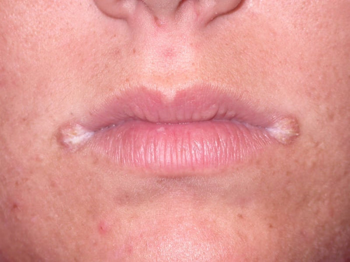
Quick Answer: A fissure (crack) at the angle of the mouth with surrounding redness and crusting is angular cheilitis. The main causes are infection — usually the yeast Candida, occasionally bacteria (Staph aureus) — and nutritional deficiencies, with saliva pooling and maceration at the mouth corners as the underlying mechanism. Work-up includes iron and vitamin B2, B6, B12 levels and swab tests where needed; treatment combines barrier creams with prescribed antifungal or antibacterial creams, and oral antifungals in selected cases. Case Snapshot: A worried patient on r/DermatologyQuestions sought help for a persistent sore at the mouth corner. On examination-style review of the images: fissuring with surrounding erythema (redness) over the left corner of the mouth, followed by crusting, with incidental Fordyce spots on the lips — a textbook presentation of angular cheilitis. The Anatomy of the Problem The angular commissures — the mouth corners — are the perfect storm for skin breakdown: they fold, they flex with every word and bite, and they collect saliva. Pooling of saliva causes physical maceration — the skin becomes waterlogged, soft, and fragile — and macerated skin splits. Once split, the warm moist crevice becomes an ideal culture plate. The Causes, Ranked Infective — the leading cause: overgrowth of Candida (a yeast) in the macerated corner; rarely bacterial, chiefly Staphylococcus aureus Nutritional: deficiencies of iron and B-vitamins (B2, B6, B12) impair the skin's repair capacity, turning a minor crack into a non-healing fissure Mechanical contributors: deep corner folds (age-related volume loss, dentures, bite changes) that increase saliva pooling The Rational Work-Up Rather than guessing, stubborn or recurrent cases get targeted tests: Blood work: iron studies and vitamin B2, B6, B12 levels — correcting a deficiency is sometimes the entire cure Swab tests from the fissure — Gram stain, KOH preparation, or culture — to confirm whether yeast, bacteria, or both are present, directing precise treatment The Treatment Ladder Barrier protection first: a bland barrier cream or lip balm applied frequently shields the corner from saliva — the mechanical fix without which everything else struggles Topical anti-infectives as prescribed: an antifungal cream such as clotrimazole for candidal cases, or an antibacterial cream such as mupirocin where bacteria are implicated Oral antifungal therapy — a dermatologist-prescribed course (such as fluconazole) reserved for extensive, resistant, or recurrent candidal cases Correct the underlying contributors: replace deficient iron/vitamins, address denture fit, and treat any associated lip-licking habit Most cases respond within one to two weeks once the right combination is in place; the commonest reason for failure is treating only the infection while the saliva-pooling and deficiency drivers continue untouched. The Fordyce Spot Footnote The incidental Fordyce spots noted on this patient's lips deserve a line of reassurance: these are normal, harmless visible oil glands, unrelated to the cheilitis, requiring no treatment — a finding, not a problem (covered fully in our dedicated Fordyce spots case). Preventing the Next Episode Once healed, a small maintenance routine keeps the corners closed: a thin film of barrier ointment at bedtime (overnight is when saliva pools longest), continued use of the bland toothpaste if it was implicated, prompt denture review where fit has changed, and finishing any prescribed deficiency correction rather than stopping when the crack heals — half-treated iron or B-vitamin deficiency simply reschedules the recurrence. Patients prone to winter episodes do well starting the bedtime ointment preemptively as the season turns. And a note on documentation worth adopting: photograph the corner during flares. Serial photos let your dermatologist distinguish true recurrence from lingering redness, and reveal patterns — always the left side, always after certain products — that no memory reliably keeps. When to See a Dermatologist See a dermatologist for any mouth-corner fissure lasting beyond two weeks, recurring repeatedly, spreading, or accompanied by oral symptoms — and before self-applying steroid creams, which temporarily soothe but feed candidal overgrowth and thin the delicate perioral skin. Recurrent cases especially merit blood work and, where product allergy is suspected, patch testing. Book a consultation here. FAQs Q: Why does my angular cheilitis keep returning on one side? A persistent local factor — an asymmetric fold, sleeping position pooling saliva on that side, or an untreated deficiency — usually explains one-sided recurrence. That's what the work-up identifies. Q: Can I just use an antifungal cream from the pharmacy? A short antifungal trial often helps candidal cases, but non-response, recurrence, or crusting warrants proper assessment — bacterial cases and deficiencies need different treatment entirely. Q: Is this a herpes cold sore? Different condition: cold sores are grouped blisters with a tingling prodrome, usually on the lip body; angular cheilitis is a fissure seated exactly in the corner fold. A dermatologist can distinguish them on sight. Q: Do dentures cause angular cheilitis? Poorly fitting dentures alter bite height and deepen the corner folds, increasing saliva pooling — a classic driver in older patients, and fixable. 📌 Case Study Reference: Publicly shared patient images and query, r/DermatologyQuestions, Reddit — "I need some help — I'm very worried…". Educational discussion only; not a final diagnosis. Clinical commentary by the Allodermis dermatology team.
Learn moreCase 25: Tiny White Bumps That Look Like Whiteheads but Never Pop — Milia Explained
Quick Answer: Tiny white bumps on the face that resemble closed comedones are often milia — small keratin-filled cysts formed when dead skin gets trapped under the surface. Unlike acne, they are not caused by oil or bacteria, so common acne treatments won't help much. Safe removal is a quick sterile procedure at a dermatologist's clinic; gentle salicylic acid exfoliation and a night-time retinoid help prevent and gradually loosen them. Case Snapshot: A user asked: "I have these closed-comedone-looking things on my face. I don't know what these are… what should I do to get rid of these?" Their images showed the classic picture: tiny, firm, pearly-white bumps scattered on the face — unchanged for months, resisting every acne product tried. What Are Milia? Milia are keratin-filled cysts — miniature pearls of trapped dead skin protein sitting just under the surface. People commonly confuse them with whiteheads, and the resemblance is genuine, but the biology is entirely different: milia are not caused by oil or bacteria. There is no clogged, active pore; there is a tiny sealed cyst with no natural exit. This single fact explains months of patient frustration — common acne treatments won't be very helpful, benzoyl peroxide does nothing, and no amount of squeezing "pops" them, because there is no opening to pop through. Attempted squeezing just bruises and marks the surrounding skin. How to Tell Milia from Whiteheads Milia: firm, pearly, uniform white; unchanged for months; won't express; no redness around them Whiteheads (closed comedones): softer, skin-coloured to white; part of a fluctuating acne pattern; respond to retinoids over weeks; occasionally inflame into pimples If a "whitehead" has sat identical and immovable for three months while your acne routine cleared everything around it — think milia. Why They Appear Common triggers include: Sun damage thickening the surface skin Skin healing after procedures, burns, blisters, or trauma — new skin can seal keratin beneath it Heavy occlusive creams in some cases, trapping dead cells Newborn milia (very common, self-resolving) and spontaneous adult milia with no trigger at all The Right Way to Remove Them Visit a dermatologist for quick removal in a sterile procedure — a tiny nick with a sterile needle or blade and gentle expression empties each milium in seconds, with no scarring when done properly. This is the definitive treatment, and a full face can be cleared in one short sitting. Gentle chemical exfoliation with salicylic acid — thins the overlying surface over weeks, helping smaller milia surface and discouraging new ones Night-time retinoid — helps by breaking down keratin and loosening the uppermost skin layer, promoting the shedding that lets milia gradually resolve and preventing recurrence What not to do: home needling (infection, scarring, and marks in Indian skin), aggressive scrubs, and comedone-extractor tools pressed onto a lesion with no opening. The Physics of Why Squeezing Fails It helps to picture the anatomy. A whitehead sits in a pore — a channel with a (blocked) exit — so pressure can, messily, express it. A milium is a sealed cyst with no channel at all: keratin wrapped in an intact capsule beneath unbroken skin. Squeezing transmits force into the surrounding tissue, bruising it and triggering pigment, while the pearl sits untouched — or ruptures sideways under the skin, inviting inflammation. The dermatologist's needle solves the geometry rather than fighting it: it creates the missing exit first, after which the contents lift out with almost no pressure. Same lesion, opposite outcome — which is the entire argument for sterile in-clinic removal over bathroom-mirror attempts. Prevention Going Forward Daily sunscreen addresses the sun-damage pathway; reviewing heavy creams and occlusive products with your dermatologist addresses another; and a maintenance retinoid keeps the surface turnover healthy. Around the eyes — a favourite milia location — choose lighter formulations and resist rich balms if you're milia-prone. When to See a Dermatologist See a dermatologist for confirmation (several other tiny facial bumps mimic milia — syringomas, flat warts, closed comedones — and each is handled differently), for sterile removal, and especially before letting any salon or home tool near lesions on the face. Book a removal appointment; combined with a chemical peel program, milia-prone skin can be kept consistently clear. FAQs Q: Will milia go away on their own? Some do, over months to a year, as skin slowly turns over. Removal is instant, safe, and usually preferred for visible facial lesions. Q: Are milia dangerous or a sign of illness? No — they are benign keratin cysts. Their only cost is cosmetic. Q: Why do I keep getting milia around my eyes? Thin eyelid skin plus rich eye creams is the usual pairing. Lighter products and a dermatologist-guided retinoid strategy reduce recurrence. Q: Can threading or facials remove milia? No — and unsterile attempts risk infection and marks. Removal belongs in a clinic. 📌 Case Study Reference: Publicly shared patient images and query, r/IndianSkincareAddicts, Reddit — "What are these on my skin?". Educational discussion only; not a final diagnosis. Clinical commentary by the Allodermis dermatology team.
Learn moreCase 24: Dark, Velvety Skin on the Neck and Underarms — Acanthosis Nigricans, the Skin's Metabolic Warning Light
Quick Answer: Darkening on the neck, underarms, or skin folds that doesn't wash off — with a thick, velvety texture and usually symmetrical distribution — is acanthosis nigricans. Its most common cause is insulin resistance, often linked to obesity or PCOS, making it an early warning sign of metabolic problems like diabetes. Treatment targets the cause first — weight management and diet — supported by lactic acid moisturisers, retinoids, chemical peels, and dermatologist-guided procedures for the skin itself. Case Snapshot: A user on r/IndianSkincareAddicts requested treatment for acanthosis nigricans — describing the classic stubborn darkening of the neck folds that no amount of scrubbing or "de-tan" remedies had shifted. What Is Acanthosis Nigricans? Acanthosis nigricans (AN) is a condition where skin becomes dark, thick, and velvety in texture, most commonly over the neck, underarms, groin, elbows, knees, and other skin folds, and is usually symmetrical. The texture is the key: this is not a surface stain but an actual change in the skin's architecture — thickened, ridged, velvet-like — which is precisely why washing, scrubbing, and bleaching creams fail. Families often mislabel it as "dirt" or poor hygiene, adding social distress to a condition that is neither. The Cause That Matters Most The most common driver is insulin resistance — the body's cells responding poorly to insulin, forcing insulin levels higher. Excess insulin stimulates skin cells in the folds to overgrow and darken. AN therefore travels with obesity and PCOS (read how hormones reshape skin and hair in our guide: PCOS Symptoms in Women), and can also appear with certain medications or, rarely, internal medical conditions — sudden, extensive, or unusual-pattern AN in particular needs thorough evaluation. The reframing every patient deserves: it's not just a skin problem — it can be an early warning sign of metabolic issues like diabetes. The neck is often where pre-diabetes first becomes visible, years before blood sugar crosses diagnostic lines. Treated as information rather than embarrassment, AN is a genuinely useful early alarm. The Work-Up A dermatologist confirms AN on examination, checks all fold sites, reviews weight history, menstrual history (for PCOS), and medications, and orders simple tests — typically fasting glucose and insulin, with hormonal panels where indicated. Ten minutes of assessment that can redirect a person's metabolic future. Treatment: Cause First, Colour Second Treating the cause: Weight management and an improved diet plan are key — AN visibly lightens as insulin resistance improves, making the neck an honest progress marker Managing PCOS or adjusting culprit medications where relevant Treating the skin: Moisturisers with lactic acid to soften and thin the velvety buildup Retinoids to normalise the overgrown skin Chemical peels and dermatologist-guided treatments, including laser toning in selected resistant cases What to avoid: aggressive scrubbing, bleach, and steroid "fairness" creams — all worsen fold skin. And beware any clinic promising instant permanent removal without addressing metabolism; the skin simply re-darkens. The Lifestyle Prescription, Made Specific "Weight management and diet" only works when translated into actions. The evidence-aligned starting set: reduce refined carbohydrates and sugary drinks — the foods that spike insulin hardest; anchor meals with protein and fibre to blunt glucose surges; walk after meals — even 10–15 minutes measurably improves post-meal insulin handling; build toward 150 minutes of weekly activity plus simple resistance work, since muscle is the body's largest glucose sink; and protect sleep, because chronic sleep debt independently worsens insulin resistance. Patients who adopt even half of this typically watch the neck lighten within months — visible feedback that few other lifestyle programs offer. Our weight management support pairs these changes with medical monitoring where needed. When to See a Dermatologist See a dermatologist for any fold darkening that is thickened or velvety, any AN appearing in childhood or spreading quickly, or simply to get the metabolic screening organised alongside skin treatment. Book an evaluation — the earlier insulin resistance is caught, the more reversible everything is, skin included. FAQs Q: Can acanthosis nigricans go away completely? It can substantially improve — sometimes near-completely — when the underlying insulin resistance is corrected, with skin treatments accelerating the cosmetic recovery. Q: Is it caused by not cleaning the neck properly? No. AN is a hormonal-metabolic skin change, not dirt. Scrubbing worsens it. Q: My child has a dark neck — should I worry? Childhood AN strongly suggests insulin resistance and deserves prompt screening; caught early, lifestyle change is remarkably effective. Q: How long until the neck lightens after starting treatment? With metabolic improvement plus topicals, expect visible change over 3–6 months. Peels can accelerate the surface component. 📌 Case Study Reference: Publicly shared patient query, r/IndianSkincareAddicts, Reddit — "Treatment of acanthosis nigricans — request". Educational discussion only; not a final diagnosis. Clinical commentary by the Allodermis dermatology team.
Learn more