

















Case Study
Case 41: Painful Recurring Boils and Scars in the Groin for Years — Hidradenitis Suppurativa (HS)
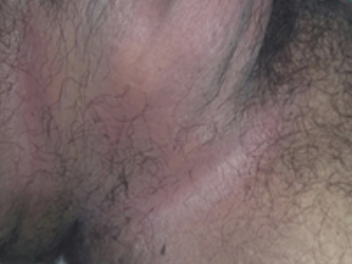
Quick Answer: Painful lumps, recurring boils with pus discharge, and scarring in the groin, armpits, buttocks, or under the breasts — persisting for years — points to Hidradenitis Suppurativa (HS), a chronic inflammatory condition of hair follicles in flexural areas. Triggers include friction, sweating, obesity, smoking, hormones, and family history. Treatment scales with severity: antibiotics and anti-inflammatory medication, biologic therapies in advanced disease, and surgery for persistent tunnels and scars. Early diagnosis matters — prompt treatment can prevent permanent scarring and dramatically improve quality of life. Case Snapshot: A patient described "tiny bumps in my pubic area" alongside a longer story: painful lumps and scars in the groin area for the past few years — the recurring boil-heal-scar cycle that defines HS, and that patients so often endure privately for years before it is finally named. What Is Hidradenitis Suppurativa? HS is a chronic inflammatory condition affecting hair follicles in flexural (fold) areas — the armpits, groin, buttocks, and under the breasts. It is emphatically not an infection caught from anywhere, and not a hygiene failure: the follicles in these friction-heavy zones become blocked and inflamed, rupture deep in the skin, and set off recurring cycles of painful inflammation. Over time patients develop: Painful boils or nodules with recurrent pus discharge Sinus tracts — tunnels under the skin connecting lesions Scarring and thickened skin in the affected folds The years-long course in this case is typical — and tragic, because every untreated year adds tunnels and scars that early treatment could have prevented. The Triggers and Risk Factors Friction and sweating in the folds Obesity — deeper folds, more friction and inflammation Smoking — one of the strongest and most modifiable associations Hormonal influences — flares often track cycles; HS typically begins after puberty Family history — a substantial genetic component The Helpful Measures Every Patient Can Start Weight management — measurably reduces flare frequency and severity Stopping smoking — among the most effective single interventions in HS Loose cotton clothing and reducing friction in affected areas — tight synthetic clothing over active sites is fuel on the fire Gentle antiseptic washes and avoiding squeezing lesions, which drives inflammation deeper Treatment: Matched to Severity Treatment depends on severity and has genuinely advanced in recent years: Antibiotics — used for their anti-inflammatory action in courses, not as one-off infection treatment Anti-inflammatory medications and hormonal approaches in suitable patients Biologic therapies in advanced cases — targeted injectable medicines that have transformed outcomes in moderate-to-severe HS Surgical procedures for persistent tunnels and scars — deroofing and excision remove disease that medicines cannot reach The strategic message of this case: early diagnosis is important because prompt treatment can help prevent permanent scarring and improve quality of life. HS is staged — and treatment started at the "recurring boils" stage protects patients from ever reaching the "tunnels and scars" stage. The Diagnosis Delay Problem HS patients average years of misdiagnosis — treated repeatedly as simple boils, "infections," or ingrown hairs — while the disease quietly stages upward. The pattern that should trigger the HS question: recurrent painful lesions, in flexural sites, on both sides, scarring over time, for months to years. If that describes you, ask the question directly at your consultation; naming the disease is the turning point. Living Between Flares Day-to-day management shrinks the disease's footprint: warm compresses ease early painful nodules; a gentle antiseptic wash in affected folds a few times weekly reduces flare frequency; loose, breathable clothing and moisture-wicking fabrics cut the friction-sweat combination; and a flare diary (dates, sites, cycle timing, stress) turns the pattern into data your dermatologist can act on — hormonal clustering, for instance, opens specific treatment doors. Keep a named clinician: HS managed by one consistent dermatologist across years outperforms scattered emergency visits for each boil. When to See a Dermatologist See a dermatologist for any recurring painful boils in the armpits, groin, buttocks, or breast folds — especially with scarring, tunnels, discharge, or a family history. HS also carries emotional weight — pain, odour anxiety, intimacy avoidance — that deserves acknowledgment and support as part of care. Book a confidential consultation here. FAQs Q: Is HS contagious or caused by poor hygiene? Neither — it is an inflammatory follicular disease with genetic and lifestyle drivers. Scrubbing harder worsens it. Q: Why do antibiotics help if it's not an infection? Certain antibiotics are used in HS primarily for their anti-inflammatory effects on the follicle, which is why they're given in structured courses. Q: Will weight loss and quitting smoking really change my HS? They are two of the highest-impact interventions available — many patients see flares shrink markedly with both. Q: Can HS be cured? There is no absolute cure yet, but modern staged treatment — lifestyle, medical, biologic, surgical — controls most cases well, and early treatment prevents the irreversible scarring stage. 📌 Case Study Reference: Publicly shared patient images and query, r/DermatologyQuestions, Reddit — "Tiny bumps appeared on my pubic area — they're…". Educational discussion only; not a final diagnosis. Clinical commentary by the Allodermis dermatology team.
Learn moreCase 40: A Round Dark Patch That Reappears in the Same Spot After Taking Medicine — Fixed Drug Eruption
Quick Answer: A well-defined, round, dusky violet patch with surrounding redness is characteristic of a Fixed Drug Eruption (FDE) — a type of drug allergy where the lesion reappears at the exact same site every time the triggering medicine is taken. Common culprits are over-the-counter painkillers (NSAIDs like diclofenac, ibuprofen, mefenamic acid), paracetamol, and some antibiotics and antifungals. The essential step is identifying and permanently avoiding the culprit drug — re-exposure causes larger, multiple, and sometimes blistering lesions. Stop the suspected medicine, see a dermatologist, and document the drug for lifelong avoidance. Case Snapshot: A patient asked, "Can anyone tell me what this is or could be — very…" sharing images of a well-defined round dusky violaceous patch with surrounding redness. The morphology alone is nearly diagnostic — and the key question a dermatologist would ask next is: what tablet did you take in the last day or two? What Is a Fixed Drug Eruption? A fixed drug eruption is a type of drug allergy where the skin lesion reappears at the exact same site whenever the triggering medicine is taken again. A population of immune cells stationed in that patch of skin has memorised the drug; each re-exposure reactivates them locally, producing the round, dusky, red-violet patch — sometimes with burning or itching — which then fades into residual dark pigmentation that marks the site between episodes. "Fixed" is the operative word: the same spot, every time, is the disease's signature and its diagnostic gift. The Usual Suspects FDE most commonly follows over-the-counter medications — which is exactly why patients rarely connect the dots themselves: Painkillers — NSAIDs such as diclofenac, ibuprofen, mefenamic acid Paracetamol Some antibiotics and antifungals, among others The everyday scenario writes itself: a headache or period pain, a familiar tablet from the chemist, and by the next day the "burn-like" patch has returned on the lip, hand, or thigh — attributed to anything but the tablet. The Typical Story Patients notice: Sudden appearance of a dark red or purple patch, usually within hours to a day of taking the drug A burning or itching sensation in the patch Recurrence at the same location after re-exposure — the tell-tale loop Residual dark pigmentation after healing, deepening with each episode Lesions can occur anywhere but commonly affect the lips, genitals, trunk, hands and feet — and genital or lip FDE is frequently misdiagnosed as infection until the drug link is found. Why Identification Is Urgent, Not Optional The most important step is identifying and avoiding the culprit medication, because the condition escalates: repeated exposure can cause larger and multiple new lesions, and blistering in severe cases — widespread blistering drug reactions are serious medical territory. Each casual re-exposure also darkens the permanent pigment stain. The management sequence is therefore clear: stop the offending (and suspected) drug now; see your dermatologist for treatment of the acute patch and the residual pigmentation; and — the step patients most often skip — document the culprit medicine for future avoidance: written in your records, saved in your phone, told to every doctor and pharmacist, with safe alternative painkillers explicitly agreed with your physician, since related drugs can cross-react. How the Culprit Is Confirmed The diagnosis is usually clinical — the fixed-site story plus the morphology. Where several drugs are candidates, dermatologists work through timing charts of intake versus flares; supervised confirmation strategies exist, but unsupervised "testing yourself" by re-taking suspects is exactly the wrong move. The healed patch's stubborn pigmentation, meanwhile, can be managed alongside our pigmentation treatments once the trigger is secured. When to See a Dermatologist See a dermatologist promptly for any round dusky patch that appeared after medication, any rash recurring at a fixed site, and urgently for multiple patches, blistering, mouth/genital erosions, or feeling unwell with a drug rash. Bring a list — or photos — of every tablet, syrup, and supplement from the preceding week; that list usually contains the answer. Book a consultation here. FAQs Q: The patch faded on its own — do I still need to find the drug? Yes. FDE always fades between exposures; the next dose brings it back bigger. Finding and banning the culprit is the actual treatment. Q: Can a medicine I've taken safely for years suddenly cause this? Yes — drug allergies can develop after years of uneventful use, which is why "but I've always taken it" doesn't exclude a drug cause. Q: Will the dark mark go away? The pigmentation fades slowly over months once re-exposures stop, and dermatologist-guided lightening treatment can help — but every new episode re-inks it. Q: Is FDE dangerous? A single classic patch is uncomfortable rather than dangerous; the risk lies in repeated exposure — multiplying, enlarging, blistering lesions — which strict avoidance entirely prevents. 📌 Case Study Reference: Publicly shared patient images and query, r/DermatologyQuestions, Reddit — "Can anyone tell me what this is or could be — very…". Educational discussion only; not a final diagnosis. Clinical commentary by the Allodermis dermatology team.
Learn moreCase 39: Recurrent Reddish Patches on the Neck and Chest Every Summer — Pityriasis Versicolor (Erythematous Variant)
Quick Answer: Non-itchy or mildly itchy reddish patches over the neck and upper trunk that recur during summers and sweating strongly suggest pityriasis versicolor (erythematous variant) — a very common superficial yeast overgrowth caused by Malassezia, a normal skin resident. It flares with hot weather, sweating, humidity, and oily skin. Management combines keeping skin dry, breathable cotton clothing, showering after sweating, and avoiding excess oily products — with antifungal shampoos, washes, or creams, and oral antifungals for extensive or recurrent cases. Case Snapshot: A patient presented with multiple reddish (erythematous) macules and patches distributed over the neck and upper trunk — only mildly itchy at most, and returning faithfully each summer with the sweat and humidity. That signature — the site, the season, the recurrence — makes pityriasis versicolor, erythematous variant, a strong clinical possibility. What Is Pityriasis Versicolor? Pityriasis (tinea) versicolor is a very common superficial fungal/yeast overgrowth caused by Malassezia — a yeast that normally lives on everyone's skin, feeding on skin oils. Under favourable conditions it multiplies and alters the skin's surface and colour in patches. "Versicolor" means exactly what it says — many colours: patches may be lighter than surrounding skin (the familiar white spots), darker, or — as in this variant — reddish/erythematous, which is why the same disease gets mistaken for allergy, eczema, or "heat rash" when it shows its red face. The Pattern That Makes the Diagnosis The clinical fingerprints in this case: Distribution over the neck and upper trunk — the oil-rich, sweat-prone zones Malassezia prefers (chest, back, shoulders complete the map) Non-itchy or only mildly itchy — a helpful separator from eczema and most allergic rashes, which itch insistently Recurrence during summers, sweating, and humidity — the yeast's growth season Fine, branny (powdery) scale on scratching the patches — a bedside sign a dermatologist elicits, and one that a simple KOH microscopy test confirms in minutes Why It Keeps Coming Back Because the organism is a permanent, normal resident of the skin, versicolor is less an infection you "catch" than a garden that overgrows in the right weather. Hot weather, sweating, humidity, and oily skin conditions are that weather — hence the honest framing every patient deserves: treatment clears each episode, and maintenance strategy prevents the next, but a single course does not banish the yeast from existence. Recurrence is a property of the condition, not a failure of the patient or the medicine. The Helpful Measures Keep the skin dry during excessive sweating — moisture is the multiplier Wear breathable cotton clothing; synthetics trap the humid microclimate the yeast loves Shower after heavy sweating or exercise — promptly, not hours later Avoid excessive oily products on the skin — body oils over the trunk feed the organism directly The Treatment Antifungal shampoos, washes, or creams form the backbone — shampoo-based treatments used as short-contact body washes cover large trunk areas efficiently — with oral antifungal medication reserved for extensive or recurrent cases under prescription. Two expectation-setting notes: first, the colour change outlasts the yeast — patches, especially pale ones, can take weeks to months to re-blend after successful treatment, so persisting colour is not persisting infection; second, for the recurrence-prone, a dermatologist can design seasonal maintenance (periodic antifungal washes through the hot months) that keeps summers clear. Using Antifungal Washes Correctly Technique decides results with wash-based treatment: apply the antifungal shampoo to the affected trunk and neck as a lather, leave it on for 5–10 minutes before rinsing (the step most patients skip), repeat on the schedule prescribed, and continue a short maintenance frequency after clearance rather than stopping the day patches fade. Treat the whole regional zone, not just visible patches — the yeast's territory always exceeds what the eye maps. When to See a Dermatologist See a dermatologist for confirmation — reddish trunk patches have several mimics (seborrheic dermatitis, pityriasis rosea, eczema) and a KOH test settles it quickly — and for extensive, frequently recurrent, or treatment-resistant patches where oral therapy and a maintenance plan are worth structuring. Post-treatment colour differences that persist can also be assessed alongside our pigmentation services. Book a consultation here. FAQs Q: Is pityriasis versicolor contagious? Practically no — the yeast lives on everyone already; the condition reflects individual skin conditions, not transmission. Q: Why are my patches red while my friend's were white? Same organism, different visible response — versicolor genuinely comes in light, dark, and red variants depending on skin tone and inflammation. Q: My patches are still visible after treatment — has it failed? Not necessarily: pigment takes weeks to months to normalise after the yeast is cleared. Fresh scale on scratching, or spread, are the true signs of active disease. Q: Can I prevent next summer's episode? Largely yes — the dryness/cotton/shower habits plus a dermatologist-planned seasonal antifungal wash routine dramatically cut recurrences. 📌 Case Study Reference: Publicly shared patient presentation, Reddit dermatology communities. Educational discussion only; not a final diagnosis. Clinical commentary by the Allodermis dermatology team.
Learn moreCase 38: Oval Scaly Patches Appearing on Both Daughters — Pityriasis Rosea, the Self-Limiting Rash That Follows a Cold
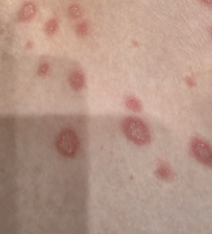
Quick Answer: Multiple oval reddish patches with fine scaling at the edges and inward peeling suggest pityriasis rosea — a common, harmless, self-limiting skin condition of children and young adults, often preceded by a recent viral illness (fever, cold, cough, or sore throat). It is thought to be linked to viral triggers, resolves on its own over several weeks, and needs only symptomatic care: moisturisers, mild topical steroids if itchy, antihistamines for itching, gentle skin care, and avoiding harsh soaps. Parents can be reassured it usually clears without scarring. Case Snapshot: A parent asked: "Any idea what this is? Both my daughters have it." Images showed multiple oval reddish patches with fine scaling at the edge, peeling inward — the distinctive "collarette" scale pattern that points to pityriasis rosea, appearing in two siblings within the same window. What Is Pityriasis Rosea? Pityriasis rosea is a common, self-limiting skin condition most often seen in children and young adults. The classic story: a few days to weeks after an ordinary upper respiratory infection — a viral fever, cold, cough, or sore throat — a rash of oval, salmon-to-reddish patches blooms across the trunk and upper limbs. Each patch carries the diagnostic signature seen in this case: a rim of fine scale at the edge that peels inward, like tissue paper attached at the border. Often (though not always) a single larger "herald patch" appears first, with the wider crop following; on the back, patches may align along skin lines in a "Christmas-tree" pattern. The Cause — Honestly Stated The exact cause is not fully established, but it is thought to be linked to viral triggers in some cases — which fits both the post-viral timing and, as here, its occasional appearance in more than one family member sharing the same seasonal infections. Importantly, the rash itself is not considered meaningfully contagious in the way infections like ringworm or scabies are — siblings developing it together reflects shared viral exposure more than rash-to-rash spread. The Course: Weeks, Then Gone This is the reassurance at the heart of the case: the condition is usually harmless and resolves on its own over several weeks — commonly 6–8, sometimes longer — and only symptomatic treatment is required. The rash may look worse before it fades; darker skin can show more prominent scaling and post-rash pigmentation that itself settles over subsequent weeks. Symptomatic Care That Helps Moisturisers — liberally, keeping patches supple and less itchy and flaky Mild topical steroids if itchy — short courses, as advised, for the more irritable patches Antihistamines for itching, especially to protect sleep Gentle skin care and avoiding harsh soaps — anything stripping worsens the scale and the itch; lukewarm baths and soap-free cleansers are the standard Loose cotton clothing; avoiding overheating and heavy sweating, which aggravate itch Parents should be reassured that this is usually not dangerous and often improves spontaneously without scarring — the sentence most families most need to hear. The Look-Alikes Worth Excluding The reason a doctor's confirmation is still worthwhile: a few conditions imitate pityriasis rosea — ringworm (usually fewer, expanding ringed patches — and antifungals treat it, unlike PR), eczema, drug-related rashes, and in sexually active age groups, secondary syphilis classically mimics PR and is checked for when the picture fits. A quick examination — occasionally with a KOH scraping to exclude fungus — settles the diagnosis and prevents weeks of wrong creams. A Simple Home Plan for the Coming Weeks For families, the practical version: moisturise the patches twice daily; give the evening antihistamine dose (as advised) on itchy nights so sleep is protected; keep baths short and lukewarm with a soap-free wash; dress the children in soft cotton; and take a weekly photo of a marked patch — watching the crop peak and then recede on camera reassures parents far better than daily close inspection, and gives the dermatologist a clean timeline if review is ever needed. When to See a Dermatologist See a dermatologist to confirm the diagnosis at the outset (especially with two children affected), if itching is severe, if the rash persists beyond about 8–10 weeks, keeps recurring, involves the face extensively, or is accompanied by feeling unwell — the cues to reconsider mimics. Book a consultation here. FAQs Q: Should my daughters stay home from school? No — pityriasis rosea is not classed as contagious, and children can attend school and play normally. Q: Will the patches leave marks? It typically clears without scarring; temporary light or dark patches in darker skin fade over weeks with moisturiser and sun sense. Q: Can they bathe and swim normally? Yes — lukewarm bathing with gentle cleansers is fine; very hot water and harsh soaps worsen itch and scaling. Q: Why did both children get it together? Shared household viral infections in the preceding weeks are the likely common trigger — a pattern occasionally seen in families. 📌 Case Study Reference: Publicly shared patient images and query, r/DermatologyQuestions, Reddit — "Any idea what this is? Both my daughters have it". Educational discussion only; not a final diagnosis. Clinical commentary by the Allodermis dermatology team.
Learn moreCase 37: Stretch Marks on the Inner Thighs After Pregnancy and Weight Change — Striae, Stages, and What Actually Helps
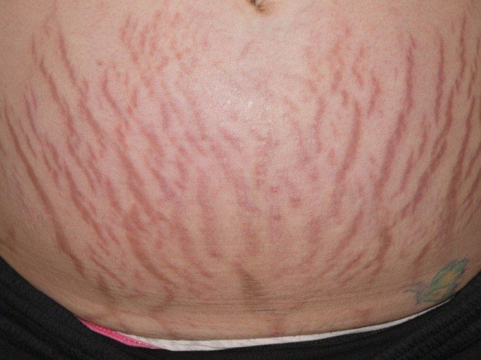
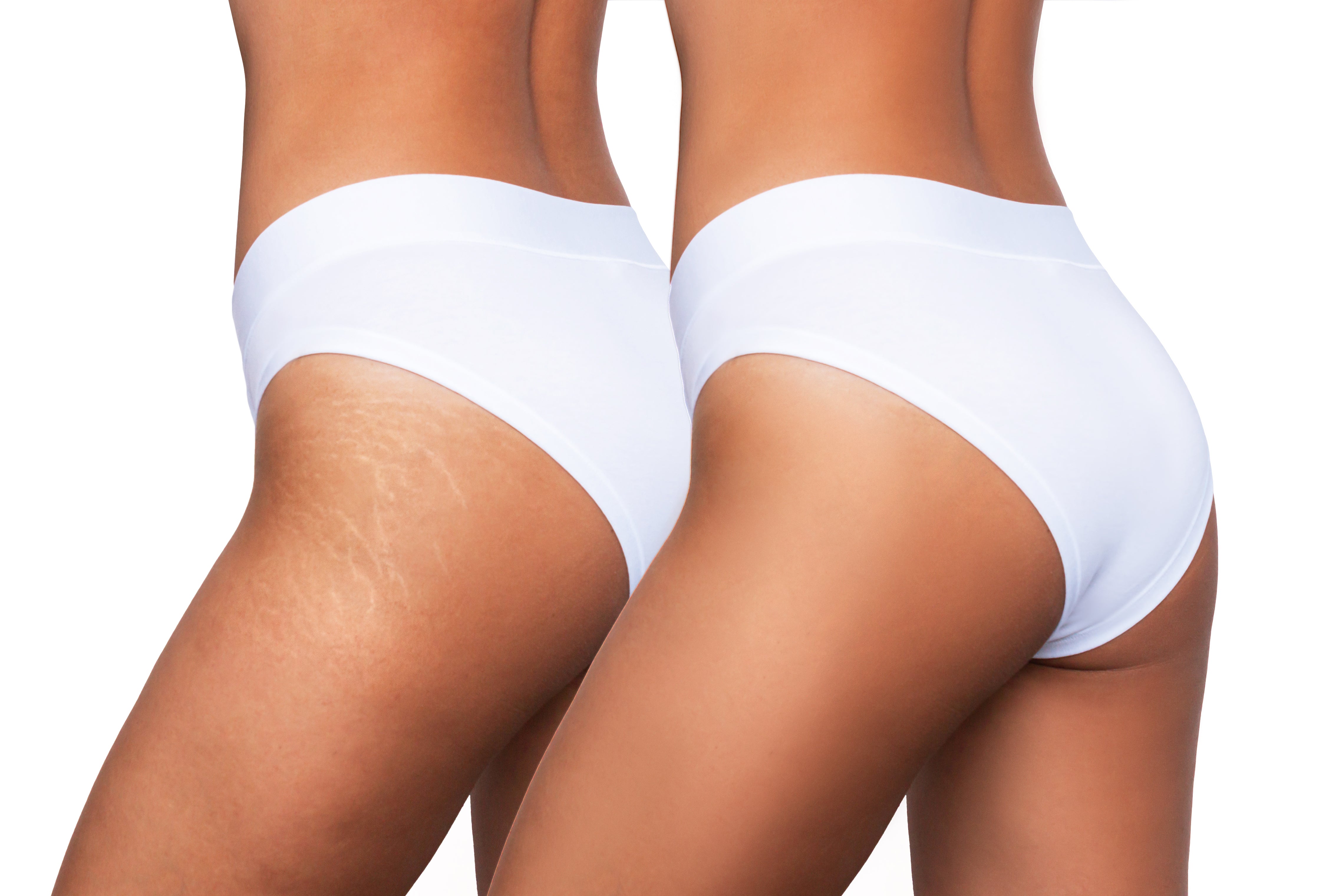
Quick Answer: Multiple linear depressed streaks on the skin are striae — stretch marks — formed when rapid skin stretching tears collagen and elastic fibres in the deeper dermis. Common causes: pregnancy, rapid weight gain or loss, hormonal conditions, adolescent growth spurts, bodybuilding, and long-term steroid use. Early red-purple marks (striae rubra) respond best to treatment; older white marks (striae alba) are harder to clear. Treatment improves texture and appearance rather than erasing marks: topical retinoids for early lesions, microneedling, fractional lasers, radiofrequency, PRP, and combination collagen-stimulating procedures. Case Snapshot: A patient shared images of "stretch marks, inner thigh, from pregnancy/weight" — multiple linear depressed streaks tracking across the inner thighs, a record of skin asked to expand faster than its fibres could accommodate. How Stretch Marks Form Striae occur when the skin stretches rapidly, leading to tearing of collagen and elastic fibres in the deeper skin layer (dermis). The surface stays intact, but the dermal scaffolding beneath ruptures in parallel lines perpendicular to the direction of stretch — which is why marks arrange themselves in those characteristic linear streams across thighs, abdomen, hips, breasts, and shoulders. The Common Causes Pregnancy — the classic setting: rapid expansion plus hormones that soften dermal fibres Rapid weight gain or weight loss Some hormonal conditions that weaken dermal structure Growth spurts during adolescence — teenage striae on thighs, hips, and backs are extremely common Bodybuilding or sudden muscle increase — shoulders and arms Long-term steroid use — oral, or potent steroid creams over-applied to thin skin (a preventable cause worth flagging) Rubra vs Alba: The Stage That Sets Your Expectations Striae age through two stages, and the stage largely determines what treatment can achieve: Striae rubra — early reddish or violaceous stretch marks: still inflamed, still biologically active, and more responsive to treatment. This is the window in which intervention pays best. Striae alba — older white or silvery marks: mature, atrophic scars of the dermis, more difficult to treat completely, though texture and visibility can still meaningfully improve. The practical takeaway: if treatment is on your wishlist, red marks now beat white marks later. The Honest Goal — and the Treatment Menu Striae are very common and medically harmless; treatment is for cosmetic confidence, and its honest aim is improving texture and appearance rather than complete removal. The evidence-aligned options: Moisturisers and barrier-repair creams — comfort, suppleness, and support during active stretching phases Topical retinoids — mainly for early lesions (striae rubra), where they measurably improve appearance (avoided during pregnancy and breastfeeding) Microneedling — collagen induction across the marks; a workhorse for Indian skin with low pigmentation risk Fractional lasers — dermal remodelling for texture and colour Radiofrequency devices and MNRF — energy-driven collagen stimulation at depth Platelet-rich plasma (PRP) — often layered with microneedling to enhance remodelling Combination collagen-stimulating protocols — in practice, pairing modalities over a series of sessions outperforms any single tool Visit our dedicated stretch marks treatment page for how these are sequenced. Prevention and Damage Limitation During pregnancy, rapid training gains, or planned weight change: keep the skin well moisturised and supple, moderate the rate of change where possible (gradual gain/loss is kinder to dermal fibres), and never use potent steroid creams on thin-skinned zones without supervision. Genetics deal the base hand — some skin marks easily regardless — but rate-of-stretch and skin condition are the levers within reach. Aftercare Around Procedures Collagen-stimulating sessions reward good habits: keep treated skin moisturised and sun-protected (fresh remodelling pigments easily in Indian skin), avoid friction from tight waistbands over healing areas for a few days, space sessions the full interval your dermatologist sets, and photograph monthly in identical lighting — striae improve too gradually for the mirror to register, but a quarter's photos show it plainly. When to See a Dermatologist See a dermatologist while marks are still red or purple to use the responsive window; for a combination-procedure plan on established white marks; or if "stretch marks" appeared without a stretching cause — widespread spontaneous striae can accompany hormonal conditions worth checking. Book a consultation here. FAQs Q: Can stretch marks be removed completely? No treatment reliably erases them; realistic outcomes are meaningful softening of texture, colour, and visibility — often substantial with combination therapy over months. Q: Do creams and oils prevent pregnancy stretch marks? Evidence for prevention is modest; consistent moisturisation supports skin comfort and elasticity, and is worth doing, but genetics and stretch rate dominate. Q: How many sessions of microneedling or laser are needed? Typically 4–8 sessions spaced weeks apart, with improvement accruing for months afterward as collagen matures. Q: When after delivery can treatment start? Topical actives and procedures are generally scheduled after pregnancy and, for several options, after breastfeeding — your dermatologist will time each safely. 📌 Case Study Reference: Publicly shared patient images and query, r/DermatologyQuestions, Reddit — "Stretch marks inner thigh from pregnancy/weight…". Educational discussion only; not a final diagnosis. Clinical commentary by the Allodermis dermatology team.
Learn moreCase 36: 'Tiny Holes' Appearing in the Skin — Anetoderma and Focal Loss of Elastic Tissue
Quick Answer: Small, soft, depressed areas that look like tiny 'holes' in the skin — sometimes feeling loose or pouch-like on touch — may represent anetoderma, a benign condition involving focal loss of elastic tissue in the skin. It can occur spontaneously or follow acne, folliculitis, trauma, chickenpox, other inflammatory skin conditions, or occasionally connective tissue disorders. These are not open pores or infections. Limited treatment options for selected cases include retinoids, fractional lasers, microneedling, and collagen-stimulating procedures — and above all, patients must stop picking or traumatising the area. Case Snapshot: A user asked: "Small holes connect in my skin — how do I fix this?" Their images showed small, soft, depressed areas giving the appearance of tiny 'holes' — with the giveaway quality that they weren't pores or pits of scar tissue, but slack little depressions in otherwise normal-looking skin. What Is Anetoderma? Anetoderma is a condition of focal loss of elastic tissue — the skin's elastin fibres, which give it snap and recoil, disappear in small discrete patches. Where elastin is lost, the skin loses its internal scaffolding tension and sags inward or outward, producing lesions that appear as: Small pouch-like or depressed areas Localized skin thinning Lesions that feel loose or herniated on touch — the classic examination sign: a fingertip pressed on the spot sinks in, as if through a small buttonhole in the skin's elastic sheet It is usually benign — a structural quirk rather than a disease process that spreads or harms. Where It Comes From Anetoderma can be: Spontaneous (primary) — arising on previously normal skin with no identifiable trigger Secondary — following previous acne, folliculitis, trauma, chickenpox, or other inflammatory skin conditions: the inflammation quietly digests the local elastic fibres, and the deficit shows itself as the depression after everything visible has healed Occasionally associated with connective tissue disorders — one reason evaluation goes beyond the skin surface in atypical, numerous, or progressive cases Not Pores, Not Infection — and Why That Matters The user's framing — "holes in my skin" — captures how alarming these look, and the first therapeutic act is correct labelling: these lesions are not true 'open pores' and not infections. They will not respond to pore products, antibacterials, or cleansing routines, and they are not something spreading through the skin. They are focal elastic-tissue deficits — closer in nature to a localized loss of the skin's rubber-band layer. Dermatologic evaluation is still useful, because similar-appearing lesions can occasionally mimic other conditions — atrophic acne scars, other atrophic disorders — and because confirming anetoderma may prompt a look for associated conditions in selected patients. The Honest Treatment Conversation Elastic tissue, once lost, is difficult for the body to rebuild — so expectations must be set honestly: treatment options are limited and help selected cases rather than erasing lesions. The options with rationale: Retinoids — supporting dermal matrix remodelling over months Fractional lasers — controlled dermal injury to stimulate new collagen and elastic tissue Microneedling and collagen-stimulating procedures (including MNRF) — the same remodelling logic used for atrophic scars, applied here to firm the depressed patches Improvement is typically partial and gradual; combinations and multiple sessions are the norm, and a dermatologist will candidly triage which lesions are worth treating. The One Instruction That Protects You Most importantly, avoid repeatedly picking or traumatising the area — trauma and inflammation are exactly what destroys elastic tissue, so worrying at the lesions can worsen texture changes and mint new ones. For secondary anetoderma, treating the driver — active acne or folliculitis — closes the factory making future lesions. When to See a Dermatologist See a dermatologist for confirmation of the diagnosis, for a considered opinion on whether procedural treatment suits your lesions, and promptly if depressions are numerous, enlarging, or accompanied by other symptoms — the scenario where associated conditions get screened. Book an evaluation here. FAQs Q: Will the holes get bigger or spread? Individual lesions are usually stable. New lesions can appear if an underlying inflammatory trigger continues — which is why treating active acne or folliculitis matters. Q: Are these the same as acne scars? They're cousins: acne scars are collagen loss, anetoderma is elastic-tissue loss — both can follow acne, both are treated with remodelling procedures, and examination tells them apart. Q: Can creams fill the depressions? No cream restores lost elastin. Retinoids modestly support remodelling; meaningful change, where achievable, comes from procedural collagen stimulation. Q: Is anetoderma dangerous? The skin lesions themselves are benign. Atypical or extensive cases are evaluated for rare associations — a check, not a cause for alarm. 📌 Case Study Reference: Publicly shared patient images and query, r/DermatologyQuestions, Reddit — "Small holes connect in my skin — how do I fix this". Educational discussion only; not a final diagnosis. Clinical commentary by the Allodermis dermatology team.
Learn moreCase 35: Severe Itching and Redness in the Groin Folds — Candidal Intertrigo, the Yeast of Warm, Moist Skin
Quick Answer: Redness and irritation of the groin folds with severe itching and burning often indicates candidal intertrigo — a yeast overgrowth in warm, moist body folds. Unlike classic ringworm, the lesions are less sharply defined and look more moist and inflamed. Triggers include sweating, tight clothing, poor drying after bathing, obesity, prolonged sitting, humidity — and notably diabetes. Management centres on keeping folds clean and completely dry, loose cotton clothing, antifungal creams or powders — and, in recurrent cases, checking for underlying diabetes. Never self-apply steroid combination creams, which worsen the infection. Case Snapshot: A patient pleaded: "Very itchy skin — please recommend some creams," sharing images of red, irritated groin folds. The appearance — moist, inflamed, less sharply bordered than ringworm — pointed to candidal intertrigo, one of the most common and most mistreated rashes of Indian summers. What Is Candidal Intertrigo? Intertrigo is inflammation of skin folds; candidal intertrigo is that inflammation driven by overgrowth of Candida yeast — a normal skin resident that flourishes when a fold turns into what it loves: warm, moist, and airless. The groin is the archetypal site; under-breast folds, armpits, and abdominal folds follow the same script. Patients complain of itching, burning, redness, soreness, and discomfort in the folds — often severe enough, as here, to dominate the day. Candida vs Ringworm: A Distinction That Changes Treatment Both are "fungal groin rashes," but they behave differently. Classic ringworm (tinea cruris) forms drier, sharply defined rings with active scaly borders. Candidal intertrigo is less sharply defined, more moist and inflamed — beefy redness deep in the fold, sometimes with small satellite spots at the edges. The distinction matters because the environments they exploit and the antifungal strategies overlap but aren't identical — and because a dermatologist seeing an atypical or resistant "fungal rash" will confirm with simple tests before escalating. Why the Fold Failed Constant friction and moisture damage the skin barrier, allowing yeast to overgrow. The trigger list is everyday life in a humid climate: Excessive sweating and humid weather Tight clothing and synthetic underwear trapping moisture Poor drying after bathing — folds that never fully dry never fully heal Obesity — deeper folds, more friction, less airflow Prolonged sitting Diabetes — the headline association: high glucose feeds yeast, and recurrent candidal intertrigo is a recognised early clue to undiagnosed diabetes The Management Plan — Counselling First The most important part of management is the environment, and dermatologists counsel it explicitly: Keep folds clean and completely dry — pat (never rub) dry after bathing; a cool-setting hairdryer on the folds is a genuinely useful trick Wear loose cotton undergarments and breathable clothing Change sweaty clothes promptly — post-gym hours in damp clothing are prime yeast time Avoid tight synthetic clothing On that foundation: antifungal creams or powders as prescribed clear the yeast, with powders doubling as moisture control for prevention. In recurrent cases, evaluation for underlying conditions like diabetes is essential — treating episode after episode while glucose runs high is bailing a leaking boat. And the emphatic warning from this case: avoid self-use of steroid combination creams. The irrational triple-combination creams sold across pharmacy counters soothe itching briefly while the steroid component feeds the infection and thins fold skin — manufacturing the chronic, modified, resistant rashes dermatologists then spend months untangling. A Daily Fold-Care Routine for the Hot Months Make prevention automatic: after every bath, pat the folds fully dry (or use a cool hairdryer for thirty seconds); dust a prescribed antifungal or plain absorbent powder before dressing; choose cotton underwear and change it — plus any sweat-soaked gym wear — the same hour it dampens; and at night, sleep in loose breathable clothing so the folds spend eight hours ventilated. Patients who ritualise these four steps through summer typically see their "annual rash" simply fail to arrive. When to See a Dermatologist See a dermatologist if the rash is severe, spreading, oozing, recurrent, or not settling within two weeks of proper antifungal care — for confirmation testing, prescription-strength treatment, and the diabetes screen recurrent cases deserve. Weight management support, where relevant, reduces recurrences at the root — see our weight management services. Book a consultation here. FAQs Q: Which cream should I buy for groin itching? Resist the pharmacy-counter combination creams. Get the rash identified first — the right plain antifungal works quickly; the wrong steroid mix creates a chronic problem. Q: Why does my rash return every summer? Heat, sweat, and humidity rebuild the yeast's habitat annually. Pre-emptive fold care — drying routines, cotton, antifungal powder in season — breaks the cycle. Q: Is this sexually transmitted? No — intertrigo is an overgrowth of resident yeast in a moist fold, not an STD, though genital rashes always deserve proper diagnosis. Q: Could this itching mean I have diabetes? One episode, no. Recurrent or stubborn candidal infections are a recognised prompt to check blood sugar — a simple test worth doing. 📌 Case Study Reference: Publicly shared patient images and query, r/DermatologyQuestions, Reddit — "Very itchy skin — pls recommend some creams or…". Educational discussion only; not a final diagnosis. Clinical commentary by the Allodermis dermatology team.
Learn moreCase 34: Small Black Spots on the Sole or Finger Pulp — Talon Noir (Black Heel), the Athlete's Harmless Mimic
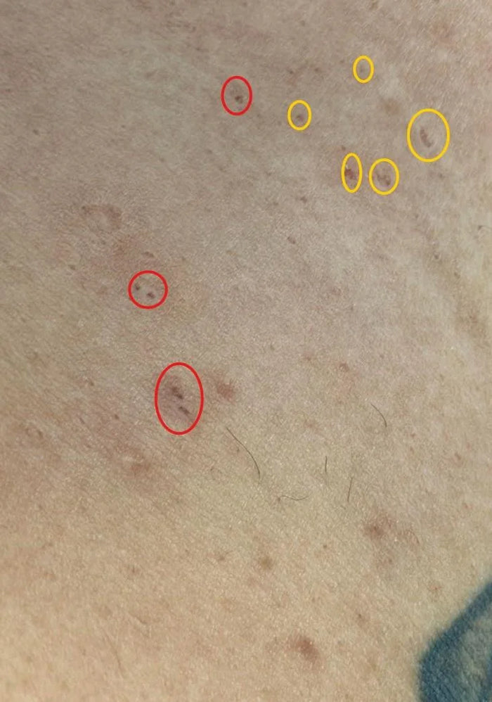
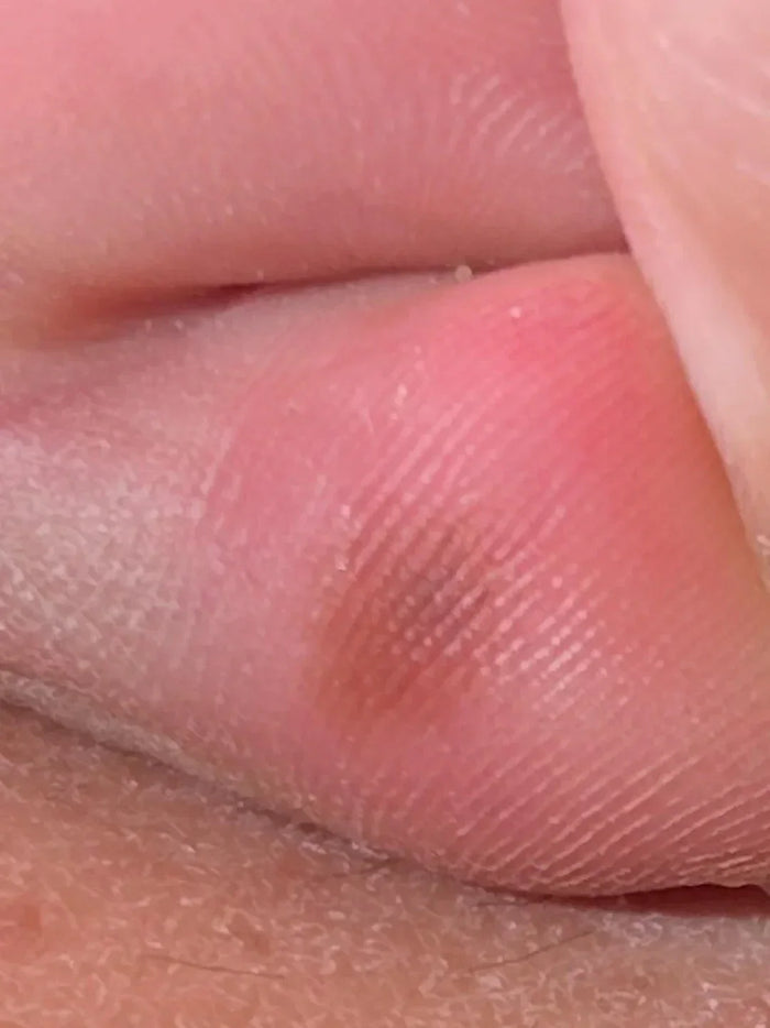
Quick Answer: Small dark brown to black spots over the sole or finger pulps are often talon noir (black heel, calcaneal petechiae) — a harmless condition caused by tiny areas of bleeding within the outer skin layer due to repeated friction or minor trauma. Common triggers are running, sports, tight footwear, and sudden increases in activity. It resolves spontaneously with reduced friction and proper footwear — but because pigmented spots on the soles must be distinguished from melanoma, dermatologists evaluate doubtful lesions carefully, often with dermoscopy. Case Snapshot: A patient asked about "pigmented spots on the sole of your foot from…" activity — images showed small dark brown to black spots over the little-finger pulp: pinpoint pigmented dots in a friction-bearing site, the classic canvas of talon noir. What Is Talon Noir? Talon noir — literally "black heel," also called calcaneal petechiae — is a harmless condition caused by tiny areas of bleeding within the outer skin layer (epidermis) due to repeated friction or minor trauma. Shearing forces rupture superficial capillaries; the escaped blood dries within the horny layer as clusters of black-red pinpoint dots. Because the blood sits in dead surface layers, the spots are painless, flat, and — crucially — temporary, shed outward as the skin renews. The Trigger List Running or sports activity — the classic setting; basketball, tennis, badminton and football players know these spots well Tight or ill-fitting footwear grinding the heel and sole margins Repeated rubbing or pressure at any site — including finger pulps in climbers, gym-goers, and racquet players, as in this case A sudden increase in physical activity — new running programs mint fresh talon noir reliably Classically described over the heel in athletes, but it equally appears as small black or brown spots on the lateral soles, ball of the foot, and finger pulps, with gradual spontaneous resolution over time once friction eases. The Serious Look-Alike — and Why Evaluation Still Matters Here is the clinical heart of this case: one important reason dermatologists evaluate such lesions carefully is to distinguish them from melanoma. Acral melanoma — melanoma of the palms and soles — presents as a pigmented macule in exactly these locations and is disproportionately important in Indian and other darker-skinned populations, where acral sites account for a larger share of melanomas. Talon noir itself is completely benign, but "black spot on the sole" is never a diagnosis to shrug at from a photo. If there is any doubt, a dermatologist may use dermoscopy — where talon noir shows its tell-tale pattern of reddish-black pebbled globules within skin ridges, and paring the surface reveals the pigment sits in (and lifts off with) the dead layer, something melanin pigment never does. The Reassuring vs Concerning Features Reassuring (talon noir): clusters of pinpoint dots rather than one growing patch; a matching activity/friction story; skin lines running undisturbed through the area; fading over weeks as activity changes. Concerning (needs review): a single enlarging pigmented patch, irregular borders or colours, a lesion unchanged or growing despite rest, any raised or ulcerated area, or pigment with no plausible friction source. Management For confirmed talon noir, treatment is refreshingly minimal: observation and follow-up; reduce the friction, repetitive trauma and pressure; wear proper footwear — cushioned, correctly sized, sport-appropriate, with moisture-wicking socks. The spots then clear spontaneously as the surface skin turns over, typically within several weeks. Nothing to excise, freeze, or bleach — and athletes can keep playing with better shoes. When to See a Dermatologist See a dermatologist for any pigmented sole or palm lesion you cannot confidently tie to friction, any spot that persists beyond 6–8 weeks despite reduced activity, or any single dark patch that grows or changes — a five-minute dermoscopy visit definitively separates the harmless athlete's mark from the lesion that must not be missed. Book a skin-lesion check here. FAQs Q: How fast does talon noir fade? Usually over 3–6 weeks with reduced friction, as the pigmented surface layer sheds. New spots appearing during heavy training are expected and equally temporary. Q: Can I scrape or pick the spots off? Don't — home paring risks wounds and confusion. In clinic, gentle paring is actually a diagnostic manoeuvre, showing the pigment lifting with the dead layer. Q: Why did this appear on my finger pulps rather than my heels? Whichever surface bears your sport's shear takes the hit — racquet grips, climbing holds, and gym bars write the same signature on the hands. Q: I have darker skin — should I worry more about sole pigmentation? Acral melanoma forms a larger proportion of melanomas in darker skin, which is exactly why any unexplained sole or palm pigmentation deserves one professional look, even though most turn out benign. 📌 Case Study Reference: Publicly shared patient images and query, r/DermatologyQuestions, Reddit — "Pigmented spots on the sole of your foot — from…". Educational discussion only; not a final diagnosis. Clinical commentary by the Allodermis dermatology team.
Learn moreCase 33: A Small Bump on the Neck With a White Center — Molluscum Contagiosum Explained
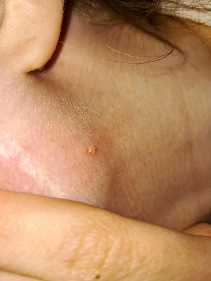
Quick Answer: A small, smooth, skin-coloured bump with whitish material visible at its center is suggestive of molluscum contagiosum — a common, harmless viral skin infection caused by a poxvirus. As the name says, it is contagious: it spreads by direct skin contact, shared towels, clothing, razors or toys, by scratching (autoinoculation), and through sexual contact in adults when lesions occur on genital skin. Treatment depends on the patient, location, and severity — options include chemical application, needling (extirpation), curettage, and cryotherapy. Case Snapshot: A user wrote: "I'm so grossed out — please help me identify this freaky bump on my neck." The image showed a small, dome-shaped, skin-coloured bump with a whitish core visible at the center — the classic single molluscum lesion, far less alarming than it felt to its owner. What Is Molluscum Contagiosum? Molluscum contagiosum is a common viral skin infection caused by a poxvirus. It produces small, firm, dome-shaped, skin-coloured to pearly bumps, often with a central dimple or visible whitish core — that core is the virus-laden material, and it is both the diagnostic giveaway and the reason scratching spreads the condition so efficiently. Lesions may be single, as here, or appear in clusters and crops. Despite the dramatic name and the disgust factor, molluscum is medically harmless — it neither scars by itself nor invades deeper tissue — and in time the immune system clears it. How It Spreads The name is honest — it is contagious, travelling by: Direct skin-to-skin contact Shared towels, clothing, razors, or toys Scratching and autoinoculation — the patient's own fingernails ferry virus from a lesion to fresh skin, planting a line or cluster of new bumps Sexual transmission in adults, when lesions occur over the genital area It is most commonly seen in children, in people in close contact with children — caregivers, nannies, parents — and in individuals with lowered immunity, in whom lesions can be more numerous and persistent. Why Location and Patient Change the Plan Treatment depends on the patient, location, and severity. A single neck lesion in a healthy adult, a face full of lesions in a toddler, and genital lesions in an adult are three different management conversations: children's lesions often warrant gentler approaches (or guided patience, since many resolve spontaneously), facial lesions demand cosmetic caution, genital lesions prompt a broader sexual-health check, and widespread molluscum in an adult can flag the need to assess immunity. The Treatment Options Chemical application — dermatologist-applied agents that provoke the lesion to resolve Extirpation (needling) — a fine sterile needle removes the central core, deflating the lesion Curettage / removal — physically scooping the lesion away; quick and definitive for accessible bumps Cryotherapy — controlled freezing with liquid nitrogen Each is a brief clinic procedure; the choice weighs speed, comfort, cosmetic site, and the patient's age. What is not on the menu: squeezing at home, which sprays infectious core material across the skin and fingernails and reliably multiplies the problem. Living With It Until It's Gone While lesions are present: don't scratch or shave over them; keep them covered where practical; use separate towels; and skip shared baths for affected children. Molluscum in swimmers and gym-goers spreads via shared surfaces and kit — a plaster over the lesion is simple insurance. Reassurance is also treatment: the bump is neither dangerous nor a hygiene verdict, and once cleared, immunity to that episode develops. When to See a Dermatologist See a dermatologist to confirm the diagnosis — several look-alikes (warts, milia, and occasionally other lesions) need different handling — and for removal, especially of facial, genital, multiplying, or inflamed lesions. Adults with genital molluscum should have a sexual-health review, and anyone with unusually widespread lesions deserves an immunity check. Book an appointment here. FAQs Q: Will molluscum go away on its own? Yes — typically over months (sometimes longer), as immunity develops. Treatment is chosen to shorten the course, limit spread, and address cosmetic or social concerns. Q: Can I go to the gym or pool with molluscum? Yes, with lesions covered and personal towels — direct lesion-to-surface contact is the risk to manage. Q: My child keeps getting new bumps — is treatment failing? New crops can surface from already-infected skin for a while; scratching accelerates this. Trimmed nails, covered lesions, and itch control help close the pipeline. Q: Does molluscum leave scars? The lesions themselves usually don't; scratched, secondarily infected, or aggressively home-treated ones can. Professional removal minimises marks. 📌 Case Study Reference: Publicly shared patient image and query, r/DermatologyQuestions, Reddit — "I'm so grossed out, please help me identify this freaky bump on my neck". Educational discussion only; not a final diagnosis. Clinical commentary by the Allodermis dermatology team.
Learn more